
SLIDE 1
Erasmus Optical Imaging Centre Zacherias (Hans) (OIC) Jansen 1595 - - PDF document
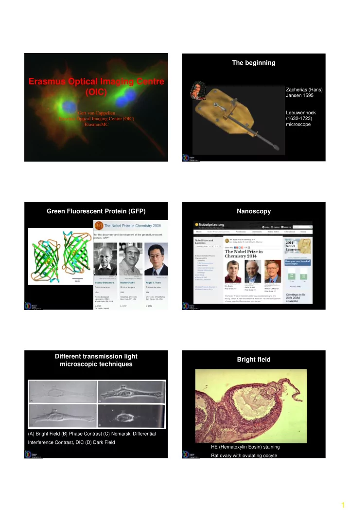
The beginning Erasmus Optical Imaging Centre Zacherias (Hans) (OIC) Jansen 1595 Gert van Cappellen Leeuwenhoek (1632-1723) Erasmus Optical Imaging Centre (OIC) microscope ErasmusMC Green Fluorescent Protein (GFP) Nanoscopy Protein
2D Stack 3D Reconstruction Volume rendering
H2b - Histone 2b protein skeleton for the DNA. One image every 10 min 14h total Hela - Henrietta Lacks (August 18, 1920 – October 4, 1951)
Distance (µm) 587 474 619 274 368 441 431 542 445 (µm) Stage I-IV I-IV I-IV I-IV I-IV V-VI VIII VIII IX-XI IX-XI Stage
Total size 4182 µm, image size 259x259µm, images rotated in advance, Andrea Enquita
Clip170-GFP Microtubule plus ends Tatiana Stepanova
movie
EXPERIMENTAL SURGICAL ONCOLOGY
Timo ten Hagen
EXPERIMENTAL SURGICAL ONCOLOGY
eNOS-GFP mouse: expression of green fluorescent protein under control of the eNOS promotor
5 minutes after tail vein injection of fluorescent microspheres, Timo ten Hagen
Axial (Z) resolution: Zmin=2*wavelength*refractive index/(Numerical aperture)2 Zmin=2*488*1.518/(1.4)2=756 nm Numerical aperture = refractive index * sin (µ) Lateral (XY) resolution: XYmin= 0.61*wavelength/Numerical aperture XYmin= 0.61*488/1.4 = 213 nm
213 nm 756 nm
µ
RAD52-PSmOrange, U2OS, Fixed cell, γ-irradiated (10 Gy), Maarten Paul
Distance (µm) 587 474 619 274 368 441 431 542 445 (µm) Stage I-IV I-IV I-IV I-IV I-IV V-VI VIII VIII IX-XI IX-XI Stage
Total size 4182 µm, image size 259x259µm, images rotated in advance, Andrea Enquita
MPL311 Zeiss LSM510NLO FCS MP, 488, 543, 633 Meta311 Zeiss LSM510Meta 266, 405, 488, 543, 633 Meta116 Zeiss LSM510Meta 488, 543, 633 Intra-Vital Meta729 Zeiss LSM510Meta 405, 488, 561, 633 LSM700_311 Zeiss LSM700, 405, 488, 555, 641 Upright LSM700_760 Zeiss LSM700, 405, 488, 555, 641 Upright Elyra Zeiss Elyra PS1, LSM780, SIM, PALM 405, 488, 561, 641, GASP 4Pi Leica 4Pi SP2, MP, 488, 561, 633 SP5_1315 Leica SP5, 405, 488, 561, 633, APD SP5_778 Leica SP5, 266, MP, 488, 561, 633, APD SP5_604 Leica SP5, 405, 488, 561, 594, 633, HyD SP5_Intravital Leica SP5, 405, 588, 561, 633, MP, OPO, NDD 4x SpinD_1454 Roper/Nikon Hamamatsu, 405, 488, 561 TIRF_1454 Roper/Nikon 405, 488, 561 LaDis2 Zeiss PALM Laserdissection microscope Perkin Elmer Opera – High content screening (CTSF)
Tsion Martijn Alex Bart Martin Johan Gert-Jan Adriaan Gert