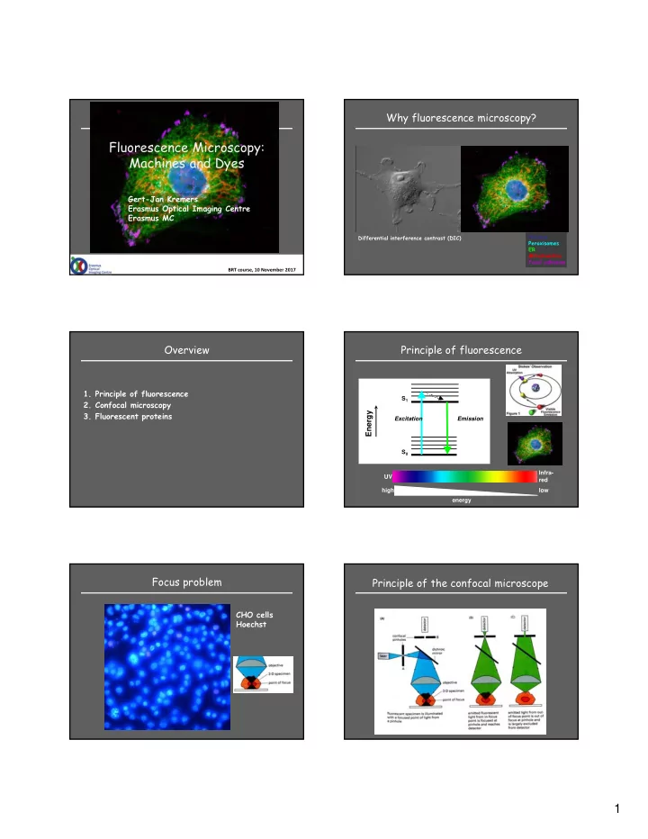

Why fluorescence microscopy? Fluorescence Microscopy: Machines and Dyes Gert-Jan Kremers Erasmus Optical Imaging Centre Erasmus MC Nucleus Differential interference contrast (DIC) Peroxisomes ER Mitochondria Focal adhesion BRT course, 10 November 2017 Overview Principle of fluorescence 1. Principle of fluorescence S 1 2. Confocal microscopy 3. Fluorescent proteins Energy Excitation Emission S 0 Infra- UV red high low energy Focus problem Principle of the confocal microscope CHO cells Hoechst 1
Optical sectioning Optical sectioning HeLa mCherry-tubulin HeLa mCherry-tubulin Pinhole at 1 airy unit Pinhole at 1 airy unit Pinhole open Pinhole open (confocal) (confocal) (widefield) (widefield) 3D scanning XY and Z laser scanning X 4 Y 3 2 1 Hela mTurquoise1_CAAX 3D scanning 3D scanning Hela mTurquoise1_CAAX MossyFiberYFP, Z. Gao, Neuroscience 2
3D scanning Fluorescent probes Blue - DAPI nucleus (direct) Green - α -tubulin (anti-body) Red - phalloidin actin (direct) MossyFiberYFP, Z. Gao, Neuroscience Fluorescent probes Fluorescent proteins – GFP gene isolated from Aq Victoria jellyfish • Organic Dyes – GFP color variants: blue, cyan, yellow – DAPI, Hoechst – First red FP from coral: DsRed – FITC, TRITC, Texas – mFruit basket of color variants Red – AlexaFluor, Cy, ATTO – Silicon rhodamine (SiR) • Quantum dots • Fluorescent proteins 27 kDa Why was GFP such a breakthrough? Genetic encoded labeling • GFP is a protein • No cofactor required • Genetic targeting: – Organism, cell, organelle – Fusion proteins – Biosensors • Low toxicity !! Live cell imaging !! http://www.conncoll.edu/ccacad/zimmer/GFP-ww/prasher.html 3
Genetic labeling Live Cell Imaging Optical highlighters Photoconvertible FPs Change in color of fluorescence after illumination with near-UV light • 3 classes – Photoactivatable FPs: dark to green – Photoswitchable FPs: dark to green (and reverse) – Photoconvertible FPs: green to red Shaner et al, JCS 2007 Photoconvertible FPs U2OS mMaple3-H2B Kremers et al (2009) Nature Methods 4
Summary • Fluorescence microscopy Fluorescence Microscopy: – Seeing is believing! – High contrast multi-color imaging Machines and Dyes • Confocal microscopy – No more blurry images > everything is in focus – Optical sectioning for 3D imaging Gert-Jan Kremers • Fluorescent proteins Erasmus Optical Imaging Centre Erasmus MC – Genetic targeting – Live cell imaging – Functional imaging Erasmus OIC BRT course, 10 November 2017 Optical Imaging Centre Rotterdam 5
Recommend
More recommend