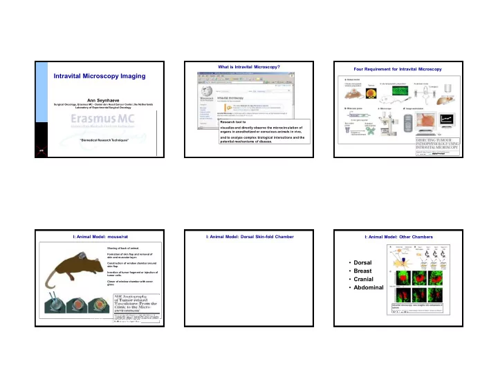

What is Intravital Microscopy? Four Requirement for Intravital Microscopy Intravital Microscopy Imaging Ann Seynhaeve Surgical Oncology, Erasmus MC - Daniel den Hoed Cancer Center, the Netherlands Laboratory of Experimental Surgical Oncology Research tool to visualize and directly observe the microcirculation of organs in anesthetized or conscious animals in vivo, and to analyze complex biological interactions and the “Biomedical Research Techniques” potential mechanisms of disease. I: Animal Model: mouse/rat I: Animal Model: Dorsal Skin-fold Chamber I: Animal Model: Other Chambers Shaving of back of animal. RAT Formation of skin flap and removal of skin and muscular layer. • Dorsal Construction of window chamber around skin flap. • Breast Insertion of tumor fragment or injection of tumor cells. • Cranial Closer of window chamber with cover glass. • Abdominal MOUSE MOUSE 1
II: Probes: Light microscopy I: Animal Model: Other Implantations II: Probes: Light Microscopy Tumor blood-flow in soft tissue sarcoma day 5 day 8 day 12 • Bones • Embryonic skin B16BL6 mouse melanoma • Lung Angiogenesis (LLC) • Heart Co-option (B16) Lewis Lung Carcinoma B16BL6 II: Probes: Fluorescent II: Probes: Fluorescent II: Probes: Fluorescent eNOS GFP Cspg4 DsRed Animals: Expression of a fluorescent protein All mTomato eNOS GFP • eNOS-GFP mouse: Green in endothelial cells • Cspg4-DsRed mouse: Red in perivascular cells Hoechst Bloodflow marker • PDGFRb-Cre x mTmG: Red in all cells, green in perivascular cells Fluorescent injectables • Drugs: Doxil, Metoxantrone, Idarubicin,… • Bloodmarkers: Albumins, Dextrans, liposomal carriers,… • Small molecules: Hoechst, DAPI,… 2
III: Microscope III: Microscope and equipment III: Microscope and equipment The complete package: Multiphoton, anesthesia unit and heated stage for long-term evaluation Most simple version: Pre-heated fixation plate for short term imaging Microscope + camera Other • Heated stage to fix the chamber Short term • Multiphoton Fixation: pre-heated metal • Confocal • Anesthesia unit plate • Fluorescent microscope Sedation: injection • High performance digital camera PC Long term • For image acquisition Fixation: plate is temperature controlled. Sedation: inhalation anesthetics. and analysis Real Time Imaging: do not miss the point Kinetics of response to therapy unknown: Histology over 5 day period. Problem: which time-point do I choose, or how many animals. - below: n= 2, 1 treatment plus control, 5 time points (Before, 4h, 24h, 48h, 72h), n=20. T Advantages of Intravital Imaging Real-time Imaging ENOUGH? C Solution: intravital microscopy does not require predefined time-point. - below: n= 2, 1 treatment plus control, continuous kinetics T C 3
Real-time Monitoring of multiple events Study the Vascular Effects by Photodynamic Therapy Real-time Monitoring of Tumor Vascular Effects 5 min eNOS GFP laser Injection of photosensitive drug Objective lens Drug 24hr microscope Rat with window and sarcoma before PDT t = 30 min (PDT) end of PDT (47 min) t = 80 min 48hr t = 4 h t = 24 h t = 48 h t = 72 h Kinetics of vessel growth Kinetics of blood flow Tissue: B16BL6 tumor Green: Endothelial cells Red: Blood marker eNOS GFP Bloodmarker Imaging dynamic processes Day 7 8 10 13 Day 14 15 16 4
Kinetics of vessel invasion Kinetics of pericyte invasion Kinetics of association and morphology eNOS GFP Cspg4 DsRed eNOS GFP Cspg4 DsRed 3D reconstruction of association and morphology Endothelial tip cell movement Imaging fast processes T=4hrs 5
Endothelial tip cell movement Filopodia movement T=60min TCR Transduced T-cell Accumulate in Tumor eNOS GFP Bloodmarker Imaging Rare Events 6
Association of pericytes at the tip cell 3D reconstruction of pericytes at the tip cell Imaging Drug Delivery Pdgfrb GFP All mTomato Monitoring Intratumoral Drug Distribution Intratumoral Drug Distribution Intracellular Drug Localization Chemotherapeutics are injected systemically Tissue: Tumor Red: Drug Injection of drug Green: Drug carrier (fluorescent) Tissue: Tumor Green: Blood marker Red: Drug carrier Question: what is the effect of certain (co)treatments on intratumoral drug distribution 7
Treatment effect on endothelial and perivascular cells Pre-treatment effects Treatment effect on endothelial and perivascular cells Tissue: Tumor Tissue: Tumor eNOS GFP Green: Endothelial cells Green: Endothelial cells eNOS GFP Red: perivascular cells Purple: Drug carrier Cspg4 DsRed Purple: Drug carrier Blue: Hoechst 2h 24h eNOS GFP Cspg4 DsRed Tissue: Tumor Green: Endothelial cells Red: perivascular cells Purple: Drug carrier 72h Hyperthermia Triggered Drug Release Hyperthermia Triggered Drug Release eNOS GFP Tissue: Tumor Green: Content Imaging of Immunological Red: Drug carrier Tissue: Tumor Processes Green: Endothelial cells Red: Drug 8
Limitations of Intravital Microscopy Intravital Imaging of Immune Processes T-cell activation by dendritic cells in the lymph node: lessons from the movies. Philippe Bousso. Nature Reviews Immunology 8, 675-684 (September 2008) 37 ° C 37 ° C • Invasive • Limited penetration depth • Movement of animal • Limited by light properties In vivo imaging of leukocyte trafficking in blood vessels and tissues. Thorsten R Mempel et al. Current Opinion in Immunology. Volume 16, Issue 4, August 2004, Pages 406 – 417 Innovation: Functional immunoimaging: the revolution continues. Nature Reviews Immunology 12, 858-864, 2012. Philippe Bousso & Hélène D. Moreau Conclusions People involved • allows real-time evaluation of processes in living Surgical Oncology Dept. Cell Biology animals. Erasmus MC Erasmus MC R. de Crom T.L.M. ten Hagen R. van Haperen T. Lu • intracellular processes can be studied. Medical University of South Carolina • correct interpretation of treatment effects. D. Haemmerich Helmholtz Zentrum München • able to study process kinetics in detail. L.H. Lindner Max Planck Institute, Munster R.H. Adams S. Adams H.M. Eilken Choose the right imaging technology for your research question. a.seynhaeve@erasmusmc.nl 9
Recommend
More recommend