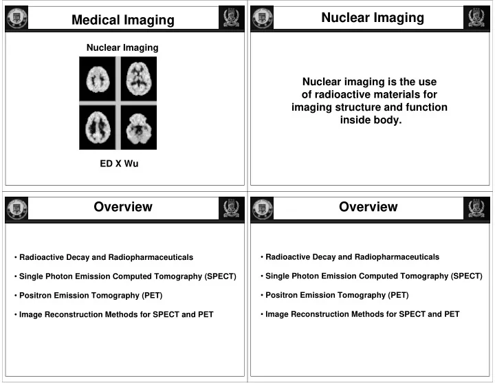
nuclear imaging medical imaging medical imaging
play

Nuclear Imaging Medical Imaging Medical Imaging Nuclear Imaging - PowerPoint PPT Presentation

Nuclear Imaging Medical Imaging Medical Imaging Nuclear Imaging Nuclear Imaging Nuclear imaging is the use of radioactive materials for imaging structure and function inside body. ED X Wu Overview Overview Radioactive Decay and


  1. Nuclear Imaging Medical Imaging Medical Imaging Nuclear Imaging Nuclear Imaging Nuclear imaging is the use of radioactive materials for imaging structure and function inside body. ED X Wu Overview Overview • Radioactive Decay and Radiopharmaceuticals • Radioactive Decay and Radiopharmaceuticals • Single Photon Emission Computed Tomography (SPECT) • Single Photon Emission Computed Tomography (SPECT) • Positron Emission Tomography (PET) • Positron Emission Tomography (PET) • Image Reconstruction Methods for SPECT and PET • Image Reconstruction Methods for SPECT and PET

  2. The Atomic Nucleus Nuclear Shell Model Analog to atomic shell model the nucleus can be Nucleus consist of protons and neutrons: described with a nuclear shell model, in which protons and neutrons occupy various possible energy states. + proton + + + + + + + + + neutron Nomenclature: A A X X or Z A := mass number (number of protons + neutrons) Z := atomic number (number of protons) Species with same Z but different A are called “isotopes.” E.g.: 64 Zn, 66 Zn, 67 Zn, 68 Zn, 70 Zn (49%, 28%, 4%, 19%, 0.6%) Stable Nuclei Decay Equation and Half-Life Nucleus is most stable when a shell is completely filled Unit of Radioactivity or Activity (to define # of with protons and neutrons. decays/disintegrations per unit of time): (Magic numbers Z or N = 2 ,8,20,28,50,82,126) (Super magic numbers Z and N = 2,8,20,28,50,82,126) Curie (Ci) & Becquerel (Bq=1/s) line of stability OR electron capture (EC) More details in: Introduction to radiological physics and radiation dosimetry By Frank Herbert Attix

  3. Decay Equation and Half-Life N = N 0 e − λ t Assignment (due in 2 weeks) N 0 := initial number of parent atoms N := parent atoms remaining after time t 1. What is the activity (in both Ci & Bq) contained in ? λ := decay constant Half-life (t 1/2 )? 1 2 = N 2. Radiocarbon dating or carbon dating is routinely used to determine the = e − λ T 1/2 age of some excavated species that lived in thousands years of ago. N 0 Please describe its principle in ~150 words. β + , β − decay, ⎛ ⎜ ⎞ => ln 1 ⎟ = − λ T 1/2 or electron capture ⎝ ⎠ 2 possible 1/2 = ln2 => T λ ( Half − Life ) Unstable Nuclei (Alpha decay) Unstable Nuclei (Alpha decay) Mass Rich Nuclei: Nucleus emits alpha particle a + + What is alpha particle?

  4. Unstable Nuclei (Alpha decay) Unstable Nuclei (Beta- decays) Neutron Rich Nuclei: Neutron becomes - β − -decay proton under emission β − -decay of electron Z increase by 1 (Z new = Z old + 1). A stays the same. β 137 −> 137 0 Cs Ba + 55 56 -1 E max is the max kinetic ? energy carried by beta- Unstable Nuclei (Beta- & Beta+ decays) Unstable Nuclei (Beta+ decays) Proton Rich Nuclei: Beta+ decay Z decrease by 1 (Z new = Z old - 1). A stays the same. In fact, complex reality to conserve energy, momentum and charges!

  5. Unstable Nuclei (Beta+ & EC decay) Proton Rich Nuclei: Eb + Neutrino: 1.57 MeV Sometimes there are two competing decay processes: - Beta+ decay (Z decreases by 1) - Capture its own electron (EC decay) Unstable Nuclei (Isometric transition) Nuclei with stable number of protons Z and neutrons N in exited state emit γ rays:

  6. Unstable Nuclei (Isometric transition) Tc −> 99 Tc + γ 99m 43 43 Single Photon Emission Overview Computed Tomography (SPECT) • Radioactive Decay and Radiopharmaceuticals • Single Photon Emission Computed Tomography (SPECT) Anger Camera • Positron Emission Tomography (PET) • Image Reconstruction Methods for SPECT and PET 99m TC

  7. Anger ( γ ) Camera Anger ( γ ) Camera Single Photon Emission Single Photon Emission Computed Tomography (SPECT) Computed Tomography (SPECT)

  8. Phantom Test SPECT Measurements SPECT Measurement SPECT Measurements SPECT Measurement unknown unknown activity & absorption activity & absorption cross-section cross-section α (x,y) vs.  µ (x,y) α (x,y) vs. µ (x,y) I( � , θ ) L 1 L max L 1 L max ⎛ ⎞ L max L max ⎜ ⎟ g ( s , θ ) = α ( x , y ) exp − µ ( x , ydl ∫ ∫ ⎟ d l scattering can degrade image ⎜ ⎝ ⎠ ⇒ use of collimators necessary! L min L 1 However spatial resolution limited to 8-15mm. problem is ill-posed! ⇒ different combinations of α and µ can yield same measurement

  9. Overview • Radioactive Decay and Radiopharmaceuticals • Single Photon Emission Computed Tomography (SPECT) • Positron Emission Tomography (PET) • Image Reconstruction Methods for SPECT and PET

  10. Overview Principles of PET In unstable nucleus (more protons than neutrons) • Positron Emission Tomography (PET) proton converts to neutron under emission of positron. • Basic Principles (Z decrease by 1 , A stays same) • Radioisotope production • PET Tracers • Clinical Examples Positron travels limited distance in tissue. http://www.crump.ucla.edu/lpp Principles of PET Positron combines with electron of nearby atom and emits two 511 keV X-ray photons that travel in opposite direction. +/- 0.25 degree

  11. Principles of PET Principles of PET Principles of PET

  12. Principles of PET Principles of PET Principles of PET - 18-FDG is a glucose analog that is widely used to image glucose metabolism - O-15 for study of blood flow or perfusion - More recently, PET tracers are employed for molecular and cellular imaging

  13. Overview • Radioactive Decay and Radiopharmaceuticals • Single Photon Emission Computed Tomography (SPECT) • Positron Emission Tomography (PET) • Image Reconstruction Methods for PET and SPECT PET Measurement PET Measurement CT Transmission Measurement CT Transmission Measurement ( measurable ( measurable unknown unknown attenuation, absorption cross-section activity cross-section coincident) µ (x,y) α (x,y) shadowgram ) X-ray source ⎛ ⎞ I = I o exp − µ ( x , y ) dl ∫ ⎜ ⎟ g ( s , θ ) = c α ( x , y ) dl ⎝ ⎠ ∫ L L ⎛ ⎞ g ≡ ln I o ⎜ ⎟ ( Signal ) ⎝ ⎠ I g ( s , θ ) = µ ( x , y ) dl ∫ L

  14. PE Tomography PE Tomography PE Tomography PE Tomography PE Tomography PE Tomography PE Tomography PE Tomography

  15. PE Tomography PE Tomography PE Tomography PE Tomography g ( s , θ ) = c α ( x , y ) dl ∫ L To obtain image from projection data use filtered backprojection algorithm, etc. Multi-ring PET Some Problems of PET Some Problems of PET (1) resolution limited to 2-5 mm because of positron mean-free path before annihilation. (2) False Coincidence Events: (a) unrelated photons arrive at same time (<20ns) (~15% of all signals) (b) one or both photons of an annihilation event are scattered (e.g. Compton scattering) (10-30% of signal) (3) Unknown photon absorption profile (2) relatively high radiation dose to patient

  16. Some Problems of PET Some Problems of PET Some Problems of PET Some Problems of PET 2D to 3D PET Configurations 2D to 3D PET Configurations Some Problems of PET Some Problems of PET Anti-scatter collimator Small detectors

  17. PET Measurement PET Measurement Transmission Measurement (as in CT) Transmission Measurement (as in CT) ( measurable ( measurable unknown unknown attenuation, absorption cross-section activity cross-section coincident) µ (x,y) α (x,y) shadowgram ) X-ray source ⎛ ⎞ I = I o exp − µ ( x , y ) dl ∫ ⎜ ⎟ g ( s , θ ) = c α ( x , y ) dl ⎝ ⎠ ∫ L L ⎛ ⎞ g ≡ ln I o ⎜ ⎟ ( Signal ) ⎝ ⎠ I g ( s , θ ) = µ ( x , y ) dl ∫ L Another approach to PET: Time-of-Flight (TOF) ? - Electronics requirement? PET vs. SPECT Or TOF electronics New Trend - CT/PET

  18. PET/CT scanner Emerging Technology - PET/CT scanner - PET/MRI scanner PET/CT scanner

Download Presentation
Download Policy: The content available on the website is offered to you 'AS IS' for your personal information and use only. It cannot be commercialized, licensed, or distributed on other websites without prior consent from the author. To download a presentation, simply click this link. If you encounter any difficulties during the download process, it's possible that the publisher has removed the file from their server.

Recommend


More recommend