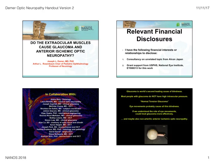

Demer Optic Neuropathy Handout Version 2 11/11/17 Relevant Financial Disclosures DO THE EXTRAOCULAR MUSCLES CAUSE GLAUCOMA AND • I have the following financial interests or ANTERIOR ISCHEMIC OPTIC relationships to disclose: NEUROPATHY? Consultancy on unrelated topic from Alcon Japan 1. Joseph L. Demer, MD, PhD Arthur L. Rosenbaum Chair of Pediatric Ophthalmology Grant support from USPHS, National Eye Institute, 2. Professor of Neurology EY008313 for this work Glaucoma is world’s second leading cause of blindness. In Collaboration With: Most people with glaucoma do NOT have high intraocular pressure. Aisha Baig - histology “Normal Tension Glaucoma” Laura Bonelli, MD - clinical optic neuropathy Joseph Caprioli, MD - clinical glaucoma Melinda Chang, MD - OCT Eye movements probably cause all this blindness. Nicolasa De Salles, MS - subject coordinator JoAnn Giaconi, MD - clinical glaucoma If we understood the role of eye movements, Vijay Gupta, PhD - mechanical engineering could treat glaucoma more effectively. Kouros Nouri-Mahdavi, MD - clinical glaucoma Bobby Lalane, MD - OCT … and maybe also non-arteritic anterior ischemic optic neuropathy. Simon Law, MD - clinical glaucoma Alan Le, BS - anatomy of optic nerve and sheath Aaron Nagiel, MD - OCT Joseph Park, BS - biomechanics and OCT Vadims Poukens, MD, PhD - histology and pathology David Sarraf - Angio OCT Steve Schwartz, MD - OCT Andrew Shin, PhD - biomechanics and OCT NANOS 2018 1
Demer Optic Neuropathy Handout Version 2 11/11/17 Take-Home Message Patient JH Progressing With Primary IOP With Open 1. In everyone, the optic nerve sheath becomes taut in adduction and 10-12 mmHg Angle supraduction, consequently tethering the globe. (Normal 10 - 21) Glaucoma 2. Modeling suggests that tethering concentrates medial rectus muscle reaction force at temporal peripapillary sclera, deforming the scleral canal and peripapillary region, and retracting the globe. 3. Medial rectus reaction force is dissipated differently in some people: A. Optic nerve and sheath elongation B. Globe translation 5. Reasons why peripapillary strain could be greater in normal tension glaucoma. A. Inner layer of optic nerve sheath stiffens with age. B. Peripapillary sclera is softer than elsewhere. 6. Repetitive strain in adduction may be a pressure-independent mechanism of optic neuropathy in glaucoma, and non-arteritic anterior ischemic optic neuropathy. 7. Extraocular muscle surgery might become an important treatment. Eye Movements Incessant Optical Coherence Tomography Adduction People make more than 180,000 saccades daily, even during sleep. Eye-head gaze shifts include eye movements averaging 30°. Saccades of up to 40-45° occur during tabletop work. Peak extraocular muscle tension is 40 gm-f for Abduction 20° saccade and 52 gm-f for 30° saccade. Peripapillary phosphenes observed during ordinary saccades suggest deformation of the optic nerve head. Model by: David A. Robinson Adduction is even greater than normal in esotropic patients. and Joel Miller NEGLECTED OPTIC NERVE Big Effect of Adduction! Chang et al. AJO, 2016. NANOS 2018 2
Demer Optic Neuropathy Handout Version 2 11/11/17 IOP 13 mmHg IOP 47 mmHg Wang, Y.X., et al., and Jonas J. B. Acute Peripapillary retinal pigment epithelium changes associated with acute intraocular pressure elevation. Ophthalmology. 122: 2022-2028, 2015. Fig. 2. Tiny Effect of IOP! Multipositional Surface Coil MRI Fiberoptic target fixation 312 micron resolution in plane 2 mm thick axial and quasi-coronal planes Digital image analysis NANOS 2018 3
Demer Optic Neuropathy Handout Version 2 11/11/17 Subjects <- Abduction Adduction -> POAG Low IOP: 19 patients Maximum IOP <20 mmHg Mean age 62 ± 10 (SD) years Mean deviation -8.2±1.2 dB Controls: 35 normals verified by examination Mean age 37± 19 yrs Age-matched control subgroup: 14 normals Mean age 63 ± 6 years (P = 0.71) All 3 groups had mean axial length 25.6 mm by MRI POAG High IOP: 2 patients ON Length ON Length Esotropia: 31 patients >>100% of ~100% of Minimum Minimum 3-D Path of Optic Nerve Temporal ON Sheath Straightest In Adduction 3-D tracking of optic neve area centroid NANOS 2018 4
Demer Optic Neuropathy Handout Version 2 11/11/17 Globe Retracts ~30° ~30° More In ON Straight Only POAG In Adduction Not Due to Age or Axial Length Effect of Adduction Still Significant After Accounting for Globe Diameter No Significant Effect of Age NANOS 2018 5
Demer Optic Neuropathy Handout Version 2 11/11/17 Finite Element Modeling Adduction -> NTG Normal Globe Translates Globe Translates Temporal More Posteriorly Mainly Nasally Scleral Temporal Temporal Stiffness Peripapillary Optic Nerve Atrophy Sheath Stiffness Nearly Universal Taut Optic Nerve Head Optic Nerve Tilts Straightens 6° Adduction Past Tethering Finite Element Analysis of Strain in Adduction Finite Element Modeling of Strain in Lamina Cribrosa During Adduction IOP 15 mm Hg Temporal -> <- Nasal IOP 15 mm Hg ICP ICP 130 mm H2O 130 mm H2O Andrew Shin, PhD NANOS 2018 6
Demer Optic Neuropathy Handout Version 2 11/11/17 Low Normal High Strain in Lamina Big Question Cribrosa Intraocular Pressure If optic nerve traction is pathologically Without Adduction -> significant, why do only some people get optic neuropathy from it? Intracranial Pressure Without Adduction -> Presumably because of individual variations in anatomy and tissue biomechanical properties. Intraocular Pressure With Adduction -> Andrew Shin, PhD 47 Retrobulbar Anatomic Dimensions Measurement (mm) Mean Std. Dev. Temporal ErrError mm Scleral Optic Nerve Diameter 3.55 0.07 Stiffness Fluid Gap Thickness 0.59 0.04 Optic Nerve Stiffer Nerve Sheath Stiffness With More Sheath Thickness 0.74 0.02 Compliant Sclera? Sheath Outer Diameter 6.16 0.12 We need human tissue biomechanical data! Nerve Sheath Stiffness Mean of 18 normal orbits. NANOS 2018 7
Demer Optic Neuropathy Handout Version 2 11/11/17 Cadaveric Studies 93 year old female Le et al. ARVO, 2017 H-7 H-8, Age 57 NANOS 2018 8
Demer Optic Neuropathy Handout Version 2 11/11/17 Biomechanics Movie by Alan Le H-8, Age 57 OCT Scanner To Linear Motor Optic Nerve Tension in Range of 10 - 60 gm To ON Sheath Force Tensile Loading Sensor Shin and Park 47 NANOS 2018 9
Demer Optic Neuropathy Handout Version 2 11/11/17 Aging Sclerosis (Hardening) of Optic Nerve Sheath Shin et al., ARVO 2017 Conclusions and Speculations Possible Options For Therapy 1. In everyone, medial rectus counterforce is transmitted to the optic nerve head in adduction by the inner layer of the temporal optic nerve sheath, stretching the optic nerve by about the same ~3%. 1. Scleral or pulley posterior fixation of the medial rectus to reduce adduction range and force. 2. The globe retracts abnormally in adduction in normal tension glaucoma, probably reflecting greater optic nerve sheath stiffness, maybe interacting 2. Combined medial and lateral rectus muscle recession to reduce with orbital connective tissues. adduction range and force. 3. The elastin content of the optic nerve sheath variably increases with age, 3. Aggressive correction of esotropia in all adults. and maybe also with normal tension glaucoma. Greater force may be required to stretch the optic nerve sheath in normal tension glaucoma, and 4. Topical (glaucoma drops) or retrobulbar prostaglandin analog therapy to this force is applied to the soft, peripapillary sclera. induce exophthalmos by orbital fat atrophy. 4. Age-related stiffening of the optic nerve sheath may be an intraocular 5. Orbital decompression by fat excision or orbital wall removal to induce pressure-independent mechanism of optic neuropathy. enophthalmos. 5. Low tension glaucoma might therefore result from repetitive strain injury 6. Other ideas? to the optic nerve head. NANOS 2018 10
Recommend
More recommend