
SLIDE 1
5/10/2013 1
Inhibition of Chondrocyte Death Following Exposure to Commonly Used Anesthetics
John G. Costouros, MD, Allison Rao, BS Tyler Johnston BA, MS, Alex Sox-Harris, PhD, R Lane Smith PhD Department of Orthopaedic Surgery Stanford University School of Medicine
Disclosures
- No disclosures relevant to the content of this
presentation.
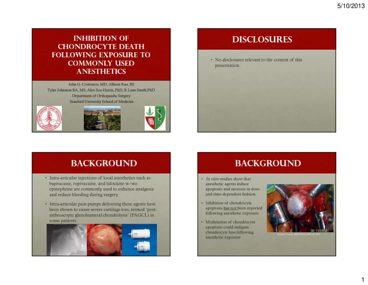
Background
- Intra-articular injections of local anesthetics such as
bupivacaine, ropivacaine, and lidocaine w/wo
epinephrine are commonly used to enhance analgesia and reduce bleeding during surgery.
- Intra-articular pain pumps delivering these agents have
been shown to cause severe cartilage loss, termed ‘post- arthroscopic glenohumeral chondrolysis’ (PAGCL) in some patients.
Background
- In vitro studies show that
anesthetic agents induce apoptosis and necrosis in dose- and time-dependent fashion.
- Inhibition of chondrocyte
apoptosis has not been reported following anesthetic exposure.
- Modulation of chondrocyte
apoptosis could mitigate chondrocyte loss following anesthetic exposure
Serrato JA et al., JBJS 93(17):e99(1-8), 2011