Active Transport by Biomolecular Motors: A New Tool for - - PowerPoint PPT Presentation

active transport by biomolecular motors a new tool for
SMART_READER_LITE
LIVE PREVIEW

Active Transport by Biomolecular Motors: A New Tool for - - PowerPoint PPT Presentation

Active Transport by Biomolecular Motors: A New Tool for Nanotechnology H. Hess, C. Brunner, J. Clemmens , K. H. Ernst, T. Nitta, S. Ramachandran, R.Tucker , D. Wu and V. Vogel Dep. of Bioengineering, University of Washington, Seattle Molecular


slide-1
SLIDE 1

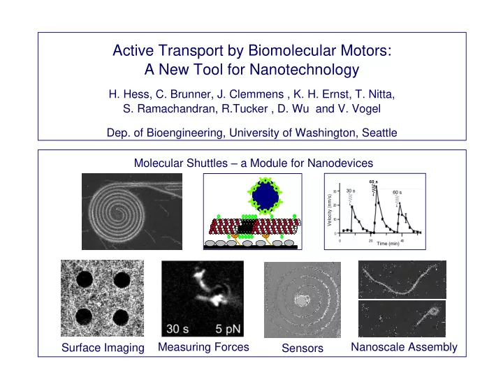
Nanoscale Assembly Molecular Shuttles – a Module for Nanodevices

Velocity (nm/s)

60 s

Surface Imaging Measuring Forces Sensors

Active Transport by Biomolecular Motors: A New Tool for Nanotechnology

  • H. Hess, C. Brunner, J. Clemmens , K. H. Ernst, T. Nitta,
  • S. Ramachandran, R.Tucker , D. Wu and V. Vogel
  • Dep. of Bioengineering, University of Washington, Seattle
slide-2
SLIDE 2

50 nm

Kinesin motor

  • N. Hirokawa, Science 279,

519 (1998)

Microtubule Kinesin

Vesicle

Movie from Vale lab, UCSF Fuel: 1 ATP per step Force: 5 pN per motor Speed: 100 steps/s of 8 nm

slide-3
SLIDE 3

Smith College, Dept. of Biology website G.A. Smith et al., PNAS, 98,3466-3470

Mitosis – coordinated movement, positional control Fast anterograde transport –

transport of vesicles (D < 100 nm) through axons ( D = 1 m) 1 m ~10 m

Motor Proteins

slide-4
SLIDE 4

Application:

Diffusion: <x 2 > = 2Dt Active transport Pressure-driven fluid flow: v ~ d2 Intracellular transport - effective diffusion limits cell size Large scale transport – cardiovascular system Self-assembly – upper limit for lateral dimensions Microfluidic devices – limit for channel diameter Mitosis Fast anterograde transport Nanofluidics Guided assembly Smart Materials

slide-5
SLIDE 5
  • J. Howard et al, Methods in Cell Biology 39 (1993) 137

Speed: 800 nm/s at 1 mM ATP

Objective slide spacers coverslip Flow in Flow out Microtubule Kinesin Casein Glass

Force: 5 pN per Kinesin Fluorescence microscope

Basic experimental setup:

Inverted motility assay

slide-6
SLIDE 6

Engineering challenges:

  • Guiding along tracks
  • Control of movement
  • Loading and unloading

Shuttle detail

Cargo Loading Cargo Delivery

Sorting

Transporting Shuttle detail Microtubule

+

Kinesin Track surface

Assembly Cargo Delivery Cargo Linkers

Shuttle system

Molecular Shuttles – a Nanoscale Transport System

slide-7
SLIDE 7

Guiding

Surface chemistry Topography Adhesion

  • f motors

Pattern layout Non-fouling surfaces Guiding channels Genetic engineering Motor concentration

Nanotechnology, 10, 232 Nano Letters, 1, 235 Langmuir, 19, 1738 Langmuir, 19, 10967 Nano Letters, 3, 1651 Lab on a Chip, 4, 83

slide-8
SLIDE 8

silicon 1 m silicon

Guiding:

Photolithography + Detergent to prevent Protein adsorption

Non-adhesive: F108 pluronic [PEO129-PPO56-PEO129] Adhesive: Bare silicon Photoresist SU8-2

Combination of surface chemistry and topography

20 m Time 50x 10 m

slide-9
SLIDE 9

Guiding:

  • H. Hess et al.: “Ratchet patterns sort molecular

shuttles”, Appl. Phys. A 75, 309 (2002) Directional sorting is accomplished by track geometry

20 m

Time 50x

slide-10
SLIDE 10

http://members.a1.net/wabweb/frames/kreuzf.htm

“Der Straßenbau“ - 1929

Woodbridge, NJ - 1928

1916 – Patent on the “clover leaf”

slide-11
SLIDE 11

Orthogonal turning path straight path

B

straight path turning path Tangential

Crossing junctions

microtubule turns around Reflector junction

  • utlet

inlet reflector arm Circular concentrator microtubule trapped in centre loop

D

Guiding: Complex track networks

  • J. Clemmens et al.: “Motor-protein “roundabouts”: Microtubules moving on kinesin-coated

tracks through engineered networks, Lab-on-a-Chip 4, 83 (2004)

10 m

100% 97% 3% 100% 67% 33%

“Gothic” “Ring” “Baroque”

slide-12
SLIDE 12

Molecular shuttles image surfaces

20 m

accessible inaccessible

Microtubule Kinesin Casein Poly- urethane

1 m

20 m 500 frames

  • bserved

in 2500 s

Nano Letters 2, 113 (2002)

slide-13
SLIDE 13

Biological Analogue: T cell trafficking

Intravital imaging of vessels (red) and cells (green) in a living lymph node

25 m

  • M. J. Miller, et al.: “Autonomous T cell trafficking examined in vivo …”, PNAS 100, 2604 (2003)

m m m

T cell perform Random Walk to sample local landscape of antigens

slide-14
SLIDE 14

1 s movie = 300 s real time

Velocity (nm/s)

Control of movement:

10 m Caged ATP is released by UV light

60 s

UV light pulses

ATP conc. reduced by added hexokinase

slide-15
SLIDE 15

Streptavidin-coated cargo Microtubules with biotin-linkers Kinesin-coated surface

Selective binding of cargo:

Streptavidin-coated cargo Microtubules with biotin-linkers Kinesin-coated surface

Selective binding of cargo:

Loading / unloading:

  • H. Hess, et al., Nano Letters, 1, 235 (2001)
slide-16
SLIDE 16

1 s movie = 50 s real time 5 m

  • H. Hess, J. Howard, and V. Vogel:

A piconewton forcemeter assembled from kinesins and microtubules,

Nano Letters, 2(10), 1113 (2002)

slide-17
SLIDE 17

A piconewton forcemeter:

slide-18
SLIDE 18

Lifetime of bionanodevices: A benchmark

  • C. Brunner, K.H. Ernst, H. Hess, V.Vogel: “Lifetime of biomolecules in hybrid nanodevices”,

Nanotechnology 15, S540 (2004)

(1) Kinesin motors are dimeric proteins undergoing large conformational changes (2) Microtubules are supramolecular assemblies of the protein tubulin, their natural equilibrium between assembly and disassembly is affected by the stabilizing anti-cancer drug taxol (used in our devices)

time [h] # of MTs per FOV Glass cell breaking disassembly

(1) Kinesin motors remain active for at least 1-2 days at room temperature. (2) Microtubules have a lifetime of ~12 hours. (3) The ATP fuel lasts for ~10 days. Microtubules are the weak link! More aggressive stabilization by chemical cross-linking required.

slide-19
SLIDE 19

Lifetime of bionanodevices: Packaging materials

  • C. Brunner, K.H. Ernst, H. Hess, V.Vogel: “Lifetime of biomolecules in hybrid nanodevices”,

Nanotechnology 15, S540 (2004)

Materials scan: Glass, PDMS, PMMA, PU, EVOH

Patterned surface: Well-characterized materials (photoresist, glass) in contact with motors Cover: Microfabricated, transparent, inert In the absence of intense illumination: Microtubule lifetimes of several hours Under illumination in the fluorescence microscope: Rapid decay for PDMS, glass cell without oxygen scavenger

O2 permeability affects compatibility

slide-20
SLIDE 20

Molecular Shuttles – a Module for Nanodevices

Velocity (nm/s)

60 s

Surface Imaging Nanoscale Forcemeter Biosensor

slide-21
SLIDE 21

Acknowledgements:

The UW molecular shuttle team: Viola Vogel Jonathon Howard (until 2002) John Clemmens, Robert Doot, Christian Brunner, Karl-Heinz Ernst Robert Tucker, Sheila Luna, Di Wu, Sujatha Ramachandran Scott Phillips The SNL motor team: Bruce C. Bunker George D. Bachand Carolyn M. Matzke, Susan B. Rivera Andrew K. Boal, Joseph M. Bauer Kinesin expression: Mike Wagenbach & Linda Wordeman Non-fouling surfaces

  • R. Lipscomb, Y. Hanein,

by plasma deposition:

  • B. Ratner, K. Böhringer

Funding: NASA, DOE-BES DARPA Biomolecular Motors Program

slide-22
SLIDE 22

Microfluidics versus Nanofluidics

500 nL sample

Baas, Microscopy Res. Techn. 48, 75

50 m Channel diameter: 50 m vs. 500 nm Sample volume: 500 nL vs. 10 fL Flow velocity: 1 mm/s vs. 1 m/s