
SLIDE 1
CENTRE FOR BIOMOLECULAR SPECTROSCOPY
Centre for Biomolecular Spectroscopy BIOMOLECULAR SPECTROSCOPY The - - PowerPoint PPT Presentation
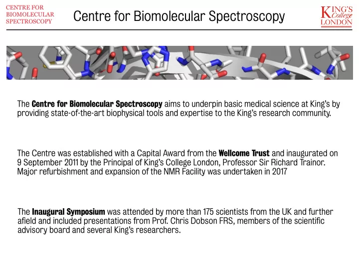
CENTRE FOR Centre for Biomolecular Spectroscopy BIOMOLECULAR SPECTROSCOPY The Centre for Biomolecular Spectroscopy aims to underpin basic medical science at Kings by providing state-of-the-art biophysical tools and expertise to the Kings
CENTRE FOR BIOMOLECULAR SPECTROSCOPY
CENTRE FOR BIOMOLECULAR SPECTROSCOPY
Isothermal titration calorimetry - ITC Surface plasmon resonance - SPR (Biacore) Optical spectroscopy - CD + High resolution protein mass spectrometry Nuclear magnetic resonance - NMR
CENTRE FOR BIOMOLECULAR SPECTROSCOPY
Interactions of human La protein with RNA - Sasi Conte (Randall) Self-association of pH responsive peptides / gene transfer - James Mason (IPS) Investigation of slow dissociation of IgE from FcεRI - Jim McDonnell & Brian Sutton (Randall)
Isothermal titration calorimetry (ITC) is the only technique that can directly measure the binding energetics of biological processes, including protein-ligand binding, protein- protein binding, DNA-protein binding, protein-carbohydrate binding, protein-lipid binding and antigen-antibody binding. ITC has the ability to determine precisely the Gibbs energy (ΔG), enthalpy (ΔH), entropy (ΔS), and heat capacity (ΔCp) changes associated with binding events.
CENTRE FOR BIOMOLECULAR SPECTROSCOPY
Reference: Velásquez-Campoy et al. (2004) Curr. Protoc. Cell Biol. 17.8.1-17.8.24
CENTRE FOR BIOMOLECULAR SPECTROSCOPY
The iTC200 instrument represents the latest generation of isothermal titration calorimetry (ITC) instruments. ITC has traditionally had very demanding sample requirements, but the iTC200 requires about ten-fold less material than the previous generation of ITC instruments: 300 μL at 50 μM : 60 μL at 500 μM GE Healthcare MicroCal iTC200
Understanding binding affjnities and interaction kinetics and thermodynamics can provide invaluable insights into the mechanisms of protein-ligand interactions.
CENTRE FOR BIOMOLECULAR SPECTROSCOPY
CENTRE FOR BIOMOLECULAR SPECTROSCOPY
Understanding binding affjnities and interaction kinetics and thermodynamics can provide invaluable insights into the mechanisms of protein-ligand interactions.
CENTRE FOR BIOMOLECULAR SPECTROSCOPY
The Biacore T100 instrument is a versatile biophysical tool for characterizing molecular interactions, enabling real-time and label-free binding studies. The exceptional sensitivity of the T100 system also allows interaction studies of low molecular weight compounds. Typically, about 1 ml of protein at 10 - 100 μg/ml is required. For the analytes, typical injection volumes would be 20 - 100 μL with concentration dependent on the expected affjnity. GE Healthcare Biacore T100
CENTRE FOR BIOMOLECULAR SPECTROSCOPY
pH titration of a linear 25 amino acid peptide
multimode spectrometers capable of measuring absorption, fluorescence and light scattering properties for ordinary, linearly and circularly polarised light for the same sample over the 170-1200 nm region.
biologicals, the study of molecular interactions and 1 ms stopped-flow kinetics.
CENTRE FOR BIOMOLECULAR SPECTROSCOPY
CENTRE FOR BIOMOLECULAR SPECTROSCOPY
Mass spectrometry is an essential tool for any protein characterization facility. The Bruker MaXis mass spectrometer ofgers ultrahigh resolution Qq-TOF technology, combining high sensitivity with exceptional mass accuracy. The instrument supports many projects in the Centre, from routine quality control of proteins undergoing structural studies, to detailed analyses of post-translational modifications and H/D exchange, as well as a number of proteomics applications. http://www.i-mass.com/guide/tutorial.html Bruker MaXis mass spectrometer
CENTRE FOR BIOMOLECULAR SPECTROSCOPY
from a useful spectroscopic technique ... ... to a powerful method for characterising macromolecular function nucleus atom polypeptide protein
biological interest.
structure, dynamics and interactions of biological macromolecules and and high throughput studies of biological fluids.
states, including lipid environments.
CENTRE FOR BIOMOLECULAR SPECTROSCOPY
CENTRE FOR BIOMOLECULAR SPECTROSCOPY
Characterisation of state of folding, oligomerisation; optimisation of domain boundaries Three-dimensional structure determination of proteins, protein domains, complexes Characterisation of protein dynamics Metal binding properties Monitoring interactions; screening of drug candidates Metabolomics Solid-state analysis
CENTRE FOR BIOMOLECULAR SPECTROSCOPY
Simple 1H NMR spectrum can indicate whether the protein is folded, pure, monomeric
CENTRE FOR BIOMOLECULAR SPECTROSCOPY
1H-15N HSQC spectra can reveal whether the protein is folded, monodisperse
7.00 8.00 9.00 10.00 110 115 120 125 130 7.00 8.00 9.00 10.00 110 115 120 125 130
CENTRE FOR BIOMOLECULAR SPECTROSCOPY
Cross-peaks are lost from the spectrum of the transcription factor interaction domain in the presence of nuclear receptor ligand binding domain. Qualitative analysis is possible even without resonance assignment.
6.50 110 115 120 125 6.50 110 115 120 125 6.50 7.00 7.50 8.00 8.50 9.00 110 115 120 125 6.50 7.00 7.50 8.00 8.50 9.00 110 115 120 125
9.0 8.0 7.0
110.0 115.0 120.0 125.0 δ15N (ppm) δ1H (ppm)
CENTRE FOR BIOMOLECULAR SPECTROSCOPY
A C-terminal extension to the canonical domain boundaries is required for successful expression and purification. N- terminal extensions are required for high quality spectra of a monodisperse protein.
PDZ1 PDZ1 PDZ1
CENTRE FOR BIOMOLECULAR SPECTROSCOPY
0.5 1 1.5
A) B) β1 β2 β3 α1 β4 α2 β5 β1 β2 β3 α1 β4 α2 β5
β1
28:-0 29:-0 3 1 :
11:7
10:7 22:91 42:32
3 9 : 3 6 4 5 : 3
55:51 56:52
3 4 : 7 7
83:79
86:82
90:22 91:22
45:30 39:36
8 5 : 8 1 8 7 : 8 3
50:47 53:50 54:50 75:72
8 2 : 7 8 23:26 3 3 :
4 : 3 4
47:28
domain boundaries is an integral part of the tertiary structure.
change in dynamics at the C-terminus.
throughout the domain may be associated with signal propagation/ allostery
J(0) (s.rad-1) J(ωN) (s.rad-1) J(ωH+ωN) (s.rad-1) x 10-9 x 10-10 x 10-11
10.0 8.0 6.0 4.0 2.0 0.0 0.0 0.0 1.0 2.0 3.0 4.0 5.0 1.0 2.0 3.0 4.0 5.0
A) B) C)
20 40 60 80 100 120
sequence
β1 β2 β3 α1 β4 α2 α5
G13 T18 F3 M-1 T30 V32 K44 S45 D49 N68 D69 G74 V81 Q85 S86 E96 L103 L105 V115 A119
CENTRE FOR BIOMOLECULAR SPECTROSCOPY
La protein is a key player in the metabolism, maturation, processing, folding and sub-cellular localisation of regulatory non- coding RNA precursors. La protects 3′ ends of newly synthesised RNA pol III transcripts from exonuclease cleavage. Highly conserved N-terminal La module (La motif + RRM1) binds with high specificity to 3′ oligoU sequences.
CENTRE FOR BIOMOLECULAR SPECTROSCOPY
Martino et al. (2012) Nucl. Acids Res., 40, 1381-1394.
In the cytoplasm, La interacts with an array of difgerent mRNAs, including the IRES domain IV of hepatitis virus C RNA.
CENTRE FOR BIOMOLECULAR SPECTROSCOPY
support the supposed secondary structure of the RNA
interaction encompasses the lower stem flanked by a single-stranded extension on either the 5′ or 3′ end
CENTRE FOR BIOMOLECULAR SPECTROSCOPY
In the cytoplasm, La interacts with an array of difgerent mRNAs, including the IRES domain IV of hepatitis virus C RNA.
CENTRE FOR BIOMOLECULAR SPECTROSCOPY
3′ oligoU
Cationic, secondary amphipathic peptides for delivery of therapeutic siRNA. Design of pH-responsive peptides, involving histidine residues, to exploit pH changes that accompany endocytosis: robust and increased delivery over non-pH responsive peptides. Increased His content was expected to promote peptide release from the peptide/nucleic acid complex, improve disordering
CENTRE FOR BIOMOLECULAR SPECTROSCOPY
Investigate:
as a function of pH correlate with results of nucleic acid delivery assay
Iacobucci et al. (2012) Biochim. Biophys. Acta, 1818, 1332-1341.
accompanied by loss of helical conformation. Greater hydrophobicity promotes self-association and leads to lower pKa.
CENTRE FOR BIOMOLECULAR SPECTROSCOPY
promoted by difgerent peptides, as a function of pH.
CENTRE FOR BIOMOLECULAR SPECTROSCOPY
Biophysical measurements provide the basis for understanding efgects of peptide sequence on nucleic acid delivery. The efgect of a more acidic pH response on peptide self-association was notable. “... strategies that seek to promote a conformational change at a less acidic pH may succeed in increasing both the effjcacy and selectivity of gene transfer”
Complementing crystallographic studies, thermodynamic analysis by SPR has shown that the interaction is entropically driven.
CENTRE FOR BIOMOLECULAR SPECTROSCOPY
The presence of the Cε2 domain alters the thermodynamic parameters from enthalpically to entropically driven, through ‘preordering’ of the intrinsically unstable Cε3 domain. Holdom et al. (2011) Nat. Struct. Mol. Biol., 18, 571-576.
CENTRE FOR BIOMOLECULAR SPECTROSCOPY
CENTRE FOR BIOMOLECULAR SPECTROSCOPY
There is no one route into the Centre! You can:
Sasi Conte; Jim McDonnell; Mark Pfuhl; Roberto Steiner; Mark Sanderson; Matthias Gautel; Brian Sutton; James Mason; Rivka Isaacson; Jane Cox; Catherine Williamson; …
There are established charges for each component of the Centre. Ask for more details. BUT - please don’t be put ofg by financial considerations - first come to see whether we can help your research.