
SLIDE 1
6/22/2013 1
Pulmonary arterio-venous fistulas
Julien I.E.Hoffman Department of Pediatrics UCSF
Pulmonary arterio-venous fistulas
- No conflicts of interest to declare
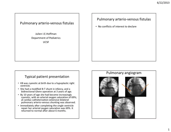
Typical patient presentation
- KB was cyanotic at birth due to a hypoplastic right
ventricle.
- She had a modified B-T shunt in infancy, and a
bidirectional Glenn operation at 3 years of age.
- By 10 years of age she had become increasingly
cyanotic, with an arterial oxygen saturation of 69%; at cardiac catheterization extensive bilateral pulmonary arterio-venous shunting was observed.
- Immediately after completing the single ventricle
repair her arterial oxygen saturation was 69%. It returned to normal after about 6 months.