
SLIDE 1
1
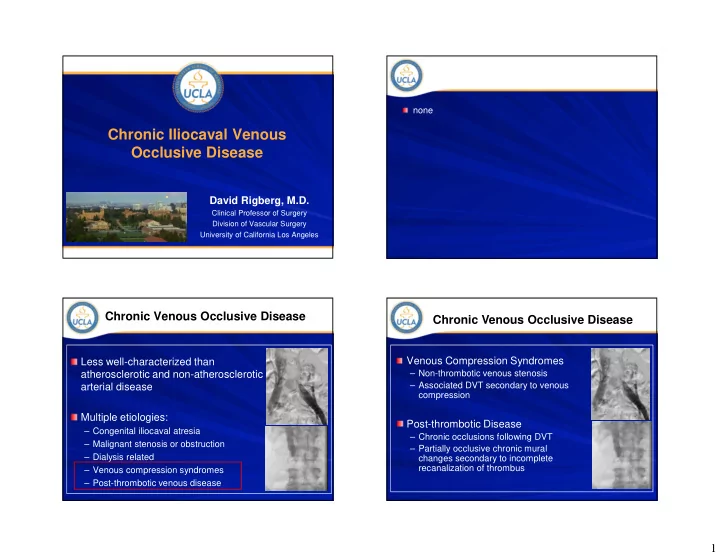
Chronic Iliocaval Venous Occlusive Disease
David Rigberg, M.D.
Clinical Professor of Surgery Division of Vascular Surgery University of California Los Angeles
Chronic Iliocaval Venous Occlusive Disease David Rigberg, M.D. - - PowerPoint PPT Presentation
none Chronic Iliocaval Venous Occlusive Disease David Rigberg, M.D. Clinical Professor of Surgery Division of Vascular Surgery University of California Los Angeles Chronic Venous Occlusive Disease Chronic Venous Occlusive Disease Venous
Clinical Professor of Surgery Division of Vascular Surgery University of California Los Angeles
Wilengberg T, LINC 2014
RCIA LCIA LCIV Compression
16 x 90 Wallstent
14 x 40 Atlas Balloon
Post Stent IVUS Immediate, 3, 6, 12 months and annually…
Incidence : up to 100,000 cases/yr (inpatient samples) < 50% complete clot lysis with anticoagulation alone 30-50% long-term risk of leg swelling and PTS Up to 15% long-term risk of venous ulcers
Pain & Venous Claudication Lipodermato- sclerosis Venous ulcers
Symptom Relief Swelling Relief Freedom from Ulcer Recurrence 528 Limbs, all with deep system reflux 69% with associated superficial or perforator vein reflux Only treatment was stenting of IVUS-determined iliac lesions 37% non-thrombotic 54% post-thrombotic 9% combined
HIP STRAIGHT HIP FLEX 90°
HIP STRAIGHT HIP FLEX 90°
Leg swelling and venous claudication / DVT Complete resolution of symptoms in most patients
History of prior DVT and IVC filter placement Technically challenging, lower success rates Dramatic symptom improvement when successful
Essential for stent sizing and positioning Post-stent assessment for residual stenosis or wall
Glycosaminoglycan (Arixtra) for 4-6 weeks in
Full antiplatelet therapy in Non-thrombotic MT
Extend stent into IVC Extend with nitinol stents into CFV if needed Aggressive lysis to improve inflow (from femoral
Thank You