Mixed Venous & Arterial Disease Mixed - - PowerPoint PPT Presentation

mixed venous arterial disease
SMART_READER_LITE
LIVE PREVIEW

Mixed Venous & Arterial Disease Mixed - - PowerPoint PPT Presentation

4/16/2016 Mixed Venous & Arterial Disease Mixed ___________________________________________________________ Arterial & Venous Venous leg ulcers are due to sustained venous hypertension. Disease: Risk factors for venous ulceration:


slide-1
SLIDE 1

4/16/2016 1

Mixed Arterial & Venous Disease:

March 16, 2016 William Tettelbach, MD, FACP, FIDSA System Medical Director of Wound & Hyperbaric Medicine Services

Can Compression Therapy Work? Mixed Venous & Arterial Disease

___________________________________________________________

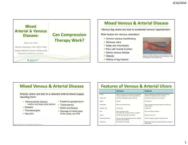
Venous leg ulcers are due to sustained venous hypertension. Risk factors for venous ulceration:

Chronic venous insufficiency Varicose veins Deep vein thrombosis Poor calf muscle function Arterio-venous fistulae Obesity History of leg fracture

Ulcer over medial malleolus of mixed arterial and venous etiology, with lipodermatosclerosis and breakdown of scar

  • ver saphenous vein harvesting site

Mixed Venous & Arterial Disease

___________________________________________________________

Arterial ulcers are due to a reduced arterial blood supply, resulting from:

Atherosclerotic disease

  • medium and large sized arteries

Diabetes Thromboangiitis Vasculitis Pyoderma gangrenosum Thalassaemia Sickle cell disease Damage of intimal layer

  • f the artery via HTN

BMJ VOLUME 332 11 FEBRUARY 2006 bmj.com

Features of Venous & Arterial Ulcers

Venous Arterial

History History of varicose veins, deep vein thrombosis, venous insufficiency or venous incompetence History suggestive of peripheral arterial disease, intermittent claudication, and/or rest pain Classic site Over the medial gaiter region of the leg Usually over the toes, foot, and ankle Edges Sloping Punched out Wound bed Often covered with slough Often covered with varying degrees of slough and necrotic tissue Exudate level Usually high Usually low Pain Pain not severe unless associated with excessive edema or infection Pain, even without infection Edema Usually associated with limb edema Edema not common Associated features Venous eczema, lipodermatosclerosis, atrophie blanche, haemosiderosis Trophic changes; gangrene may be present Treatment Compression is mainstay Appropriate surgery for arterial insufficiency; drugs of limited value

slide-2
SLIDE 2

4/16/2016 2

Compression classes of compression hosiery used in several countries.

(Values in mmHg, 1 mmHg = 1,333 hPa)

The values indicate the compression exerted by the hosiery at a hypothetical cylindrical ankle.

USA United Kingdom France Germany

15 - 20 (moderate) 14 - 17 (light) 10 - 15 18 - 21 (light) 20 - 30 (firm) 18 - 24 (medium) 15 - 20 23 - 32 (medium) 30 - 40 (extra firm) 25 - 35 (strong) 20 - 36 34 - 46 (strong) 40+ > 36 > 49 (very strong)

Eur J Vasc Endovasc Surg Vol 35, April 2008

Compression Therapy

___________________________________________________________

ABI Perfusion Status

> 1.3 Elevated, incompressible vessels 1.0 - 1.3 Normal ≤ 0.9 Lower extremity arterial disease suspected ≤ 0.6 to 0.8 Borderline ≤ 0.5 Severe ischemia < 0.4 or a toe pressure < 30 mmHg Critical Ischemia, Limb Threatening

Arterial Brachial Index

___________________________________________________________

Arterial Brachial Index

___________________________________________________________

CONTRAINDICATIONS for ABI:

Excruciating pain in lower legs/feet. Deep vein thrombosis (DVT), which could lead to dislodgement of the thrombosis, where referral would be indicated for a duplex ultrasound test. Severe pain associated with lower extremity wound(s).

  • 1. Grenon SM, Gagnon J, Hslang J. Ankle-brachial index for assessment of peripheral arterial disease. N Engl J Med. 2009;361:e40.

1

30 - 40 mmHg wrap (resting pressure)

  • Used on patients with an ABI equal to 0.8 or greater.

20 - 30 mmHg wrap (resting pressure)

  • Used on patients with an ABI equal to 0.5 or greater.

Compression Therapy

___________________________________________________________

Indications for a coban 2 layer compression system:

Junger M, Haase H, Ladwig A, Schwenke L, Bichel J, Schuren J. Compression therapy in patients with peripheral arterial occlusive disease: A prospective clinical study with the 3M Coban 2 Layer Lite Compression System for ABPI > 0.5. 2010.

slide-3
SLIDE 3

4/16/2016 3

Mixed Venous & Arterial Disease

___________________________________________________________

In patients with mixed ulceration, an ankle-brachial pressure index >0.5 and an absolute ankle pressure of >60 mmHg, inelastic compression of up to 40 mmHg does not impede arterial perfusion but may lead to a normalization of the highly reduced venous pumping function. Such bandages are therefore recommended in combination with walking exercises as the basic conservative management for patients with mixed leg ulcers.

Mosti G, Iabicella ML, Partsch H. Compression therapy in mixed ulcers increases venous output and arterial perfusion. Vasc Surg 2011;55:122–8.

Compression Therapy

___________________________________________________________

Absolute CONTRAINDICATIONS for application of compression:

A florid infection (e.g., phlegmon, abscess) Advanced peripheral arterial occlusive disease

  • ABI is < 0.5 mmHg or a toe pressure < 30 mmHg

Acute decompensated heart failure Phlegmasia coerulea dolens

  • extensive thrombotic occlusion of the major and the collateral veins
  • 1. EWMA JOURNAL 2013 VOL 13 NO 2

1

Once ulcer has healed, goal is to prevent recurrence:

Wearing compression stockings Skin care Leg elevation Calf exercises Optimizing diet

Mixed Venous & Arterial Disease

___________________________________________________________

Questions?

Thank you for your time