African trypanosome Life Cycle epimastigote salivary gland - - PDF document

african trypanosome life cycle
SMART_READER_LITE
LIVE PREVIEW

African trypanosome Life Cycle epimastigote salivary gland - - PDF document

African trypanosome Life Cycle epimastigote salivary gland metacyclic trypomastigote non-dividing infection for vertebrate host procyclic trypomastigote salivary gland fly midgut dividing form metacyclic replication inflamation (chancre)


slide-1
SLIDE 1

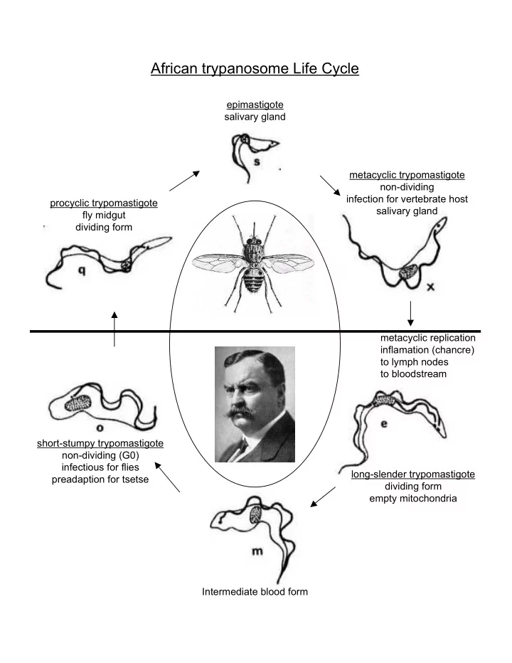
procyclic trypomastigote fly midgut dividing form epimastigote salivary gland metacyclic trypomastigote non-dividing infection for vertebrate host salivary gland short-stumpy trypomastigote non-dividing (G0) infectious for flies preadaption for tsetse long-slender trypomastigote dividing form empty mitochondria metacyclic replication inflamation (chancre) to lymph nodes to bloodstream Intermediate blood form

African trypanosome Life Cycle

slide-2
SLIDE 2

Stages of T. brucei Bloodstream Infection Stage 1 – Long-slender trypomastigotes actively divide Stage 2 – At high cell densities, long-slenders differentiate into short-stumpys (able to differentiate further into procyclics) Stage 3 – Mostly short-stumpys, immune clearance of long- slenders Stage 4 – Immune clearance of short-stumpys Stage 5 – Next proliferation of long-slenders which can evade host immune response (How? Antigenic Variation)

1 4 2 3 5

slide-3
SLIDE 3

Tseste bite Trypanosomal chancre Asymptomatic BLOOD Fever, malaise, rash alternating asymptomatic

flu-like symptoms

LYMPHATICS CNS DEATH PRIMARY BLOOD STAGE LATE STAGE Metacyclic Subcutaneous Itching 2-3 d Swelling 6 d Long slender Short stumpy Long slender

Meningoencephalitis Cerebral edema Hemorrhage Pericarditis Coma pneumonia

Pathogenesis of African Trypanosomiasis

slide-4
SLIDE 4

Invention of DDT Widespread spraying campaign Drug treatment: Painful, expensive, and potentially ineffective

slide-5
SLIDE 5

Variant Surface Glycoprotein

  • Episodes of relapsing parasitemias
  • Variant Antigenic Type
  • switching rate 1:10,000 to 1:1,000,000 per division
  • 300-1000 VSG genes per tryp.
  • 5-10% of the genome
  • serodemes – lineages of antigenically different tryps. w/ common ancestor
  • isogenic mutants – differ by the expression/function of single gene (VSG)
  • VSG cDNA - N-term. cleaved during membrane transport

C-term. removed, GPI anchor

  • One VSG gene expressed per tryp., no mixing
  • Variation of expressed VSG is random
  • ~20 different ES (10-20 for metacyclic VSG expression)
slide-6
SLIDE 6

Leishmania major chromosome 1 gene

  • rganization: polycistronic transcription
  • No transcriptional regulation of mRNAs
  • Adjacent genes are frequently not related in function
  • trans-splicing resolves individual mRNAs from

polycistronic transcripts

Switch

slide-7
SLIDE 7

+ AAAAAA pre-mRNA mRNA m7G cap intron exon exon RNA Pol II

Spliced Leader and the Processing of Polycistronic pre-mRNAs into discreet messages

slide-8
SLIDE 8

Immunoprecipitation

Epitope Tagging

Adding a sequence coding a peptide fragment that you already have Ab for.Protein C, HA, c-Myc, Strep, Flag, BB2….the list goes on and on. RNA Pol I RNA Pol I Phleo Phleo Prot C RNA Pol I Prot C Phleo Phleo Now we can easily IP and detect RNA Pol I Cell extract from: wild type wild type RNA Pol I tag RNA Pol I tag Anti-RNA pol I Anti-Protein C

Protein A binds the Fc region of Abs

slide-9
SLIDE 9

Primer Extension – measures RNA quantity

Invitro trancription

Lyse cells Extract/purify Trascriptionally active fraction Plasmid with the target gene DNA rNTPs, buffer etc. Incubate for a while Extract RNA and analyze

slide-10
SLIDE 10

Ab binding is Ca++ dependent

WT Prot C WT Prot C One of these things is not like the

  • ther…

Immunodepleting the extract abolishes Pol I transcription (Gpeet, rRNA, VSG) but does not change Pol II transcription(SL RNA) Compare the + CA with the - CA

Western blot detects the presence of RPA1

Invitro transcription This shows that the extract is clear of tagged Pol I Immuno depletion

Primer extension to detect invitro trxn RNA

The Gunzl RNA POL I experiment

slide-11
SLIDE 11
slide-12
SLIDE 12

Progression VSG switching

Early: Full genes stored generally in the minichromosomes Late: Mosaic VSG’s made from multiple recombined pieces of sub telomeric psuedo genes

slide-13
SLIDE 13

Trypanosome Lytic Factor 1

High density lipoprotein complex (phospholipids, cholesterol, cholesterol ester, Apo-proteins) ApoL-1 is the lytic factor of TLF1 TLF1 complex is endocytosed by the trypanosome There is a fusion with the lysosome, swelling of lysosome, cell lysis

slide-14
SLIDE 14

Serum Resistance-Associated Gene (SRA)

  • occurs in natural human serum (NHS)
  • structure similar to N-term. fold of VSG
  • T.brucei lacks the SRA gene – lysed in humans
  • addition of SRA to T.brucei confers resistance to lysis

control control + NHS SRA SRA + NHS

slide-15
SLIDE 15

a, Incubation of ETat 1.2S with differently treated NHS (SRA-ft, L61P/I62P- ft, aApoL-ft indicate flow-through fractions from SRA–, L61P/I62P SRA– and anti-apoL-I–Sepharose, respectively; aApoL-el, eluate of the fraction bound to anti-apoL-I–Sepharose). b, Incubation of ETat 1.2S in either SRA-ft or FCS supplemented with recombinant apoL-I (C-del, lacking the C-terminal 343–398 peptide) or with the equivalent fraction from control CHO cells (ctl). NHS = Normal Human Serum (lytic activity)) FCS=fetal calf serum (no lytic activity)

These two columns bind ApoL-I, thus the flow through (FT) will be depleted for that protein. These depleted serums have lost lytic activity Adding Apol 1 (Apol-1 WT) back to either Apol-1 depleted serum (top box)

  • r to FCS (bottom box)

confers lytic activity to these serums

slide-16
SLIDE 16

SRA Protein binds to apoL-1

Remember: ApoL-1 in NHS causes tryp. lysis SRA confers resistance to lysis Depletion of ApoL-1 (IP) from NHS removes lytic activity Addition of ApoL-1 to depleted serum restores lysis