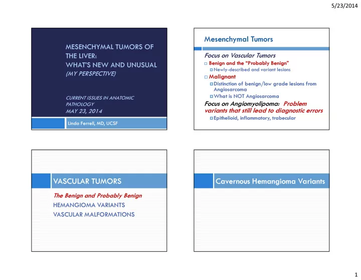
SLIDE 1 5/23/2014 1
MESENCHYMAL TUMORS OF THE LIVER: WHAT’S NEW AND UNUSUAL
(MY PERSPECTIVE)
CURRENT ISSUES IN ANATOMIC PATHOLOGY
MAY 23, 2014
Linda Ferrell, MD, UCSF
Mesenchymal Tumors
Focus on Vascular Tumors
Benign and the “Probably Benign” Newly-described and variant lesions
Malignant Distinction of benign/low grade lesions from
Angiosarcoma
What is NOT Angiosarcoma
Focus on Angiomyolipoma: Problem variants that still lead to diagnostic errors
Epithelioid, inflammatory, trabecular
The Benign and Probably Benign HEMANGIOMA VARIANTS VASCULAR MALFORMATIONS
VASCULAR TUMORS Cavernous Hemangioma Variants
SLIDE 2 5/23/2014 2
Cavernous Hemangioma (CH)
Not true arterial
architecture
No organized muscle bundles No elastic laminas Not capillary-like
Cavernous Hemangioma
Incidental (Autopsy finding Giant CH, with organized thrombosis and sclerosis
Sclerosis within Cavernous Hemangioma
Sclerosis of thrombosed, ischemic zones with scar formation. “Neo-vessels” Recanalized channels
Cavernous Hemangioma:
What is often “not seen”….
Hemangioma-like vessels (HLV) in
adjacent liver commonly seen with giant CH
Ref: Kim GE, Thung SN, Tsui WMS, Ferrell LD. Hepatic
Cavernous Hemangioma: Under-Recognized Associated Histologic Features. Liver Int'l, 26:334-38, 2006.
Low mitotic/proliferative rate <5% Present in almost 80% (16/19) of CH >5 cm Retain composition of vascular walls in CH
SLIDE 3
5/23/2014 3
Giant Cavernous Hemangioma
Cavernous Hemangioma-like vessels in adjacent liver Giant Cavernous Hemangioma
Explant, right lobe 38 yr old woman, in liver failure.
Giant Cavernous Hemangioma
Left Lobe: Smaller, irregularly shaped CHs and transitional areas with HLVs admixed with liver
SLIDE 4 5/23/2014 4
Giant Cavernous Hemangioma
Right Lobe CH Left lobe HLV Lesion extending into hilum around arteries, nerves and ducts Omental Lesion
Artery Nerve Duct
“Metatastatic” and “Invasive” Cavernous Hemangioma
Cavernous Hemangioma Variant
Diagnoses: Giant Cavernous Hemangioma and Cavernous Hemangiomatosis
CH-like vessels throughout liver, involving
hilum
Lung, spleen, omentum involved with CH-like
lesions
Problematic cavernous hemangioma variants and other benign mimics: A Mattis, S Fischer, H Makhlouf, W Tsui, S Cho, L Ferrell. Poster at USCAP Mar 2010, published Mod Pathol Supple 1, 2010.
Vascular Malformations
Hereditary Hemorrhagic Telangiectasia
(HHT) arterial-venous malformations
also known as Osler-Weber-Rendu Other Arterial and Venous Malformations
with similar features
(may or may not be HHT)
SLIDE 5 5/23/2014 5
Vascular Malformations
Contributors and co-authors of 2 abstracts:
Cho S, Paradis V, Pai R, Bioulac-Sage P
, Alves V, Souza T, Makhlouf H, Schirmacher P , Evason K, Ferrell L. Histopathologic Features of Extensive Hepatic Vascular
- Malformations. Mod Path 23 (Supple 1):352A, 2010.
Cho S, Wanless I, Sempoux C, Paradis V, Pai R, Thung S,
Bioulac-Sage P , Balabaud C, Makhlouf H, Schirmacher P Alves V, Souza T, Evason K, Ferrell L. FNH-Like Lesions and Glutamine Synthetase Expression in the Liver in Hereditary Hemorrhagic
- Telangiectasia. Mod Path, 24 (Supple 1):358A, 2011.
Vascular Malformations
Spectrum: Early, mild To Late, severe
Early or mild lesions can look much different than advanced or severe lesions probably primarily due to thrombosis and ischemic effects Vascular Malformations:
Early Lesions or Mild Involvement
Periportal fibrosis, Elastochrome stain Periductal fibrosis (as early ischemic lesion)
Vascular Malformations:
More Severe or Advanced Lesions
Extension of lesions into sinusoids Thrombosis within vessels and sinusoids
SLIDE 6 5/23/2014 6
Vascular Malformations Severe sinusoidal changes
Vascular Malformations:
More Severe or Advanced Lesions
Hemangioma-like changes, extensive sinusoidal dilation Cavernous hemangioma-like transformationn
Small Vessel Hemangioma
Rare Newly described
Small vascular channels with thin
walls
Bland endothelial cells with low
proliferative rate <10% (CH <5%)
Intermediate tumor cell density Irregular “infiltrative” growth
pattern at border
abnormal liver architecture mimics HCC scaffolding effect mimics angiosarcoma
Small Vessel Hemangioma
Small channels, thin walls, bland nuclei Only focal fibrotic areas (no wide walls as in CH)
SLIDE 7 5/23/2014 7
Small Vessel Hemangioma
Small channels with thin walls, no
Low Mib1 (Ki-67) rate
Small Vessel Hemangioma
Center of lesion, bland endothelial cells Edge of lesion, with altered cell plate width
Small Vessel Hemangioma
Edge of lesion, trichrome Edge of lesion, reticulin
Small Vessel Hemangioma
Small vessel hepatic hemangioma (SVH): Exact outcome not
definitive, so now recommending excision and followup.
Differentiation from angiosarcoma: AS has higher
proliferative rate (>15%) and subset + for P53 and GLUT1, but negative in small vessel hemangioma References
Gill R, Sempoux C, Makhlouf H, Thung S, Alves V, Ferrell L.
Small Vessel Hepatic Hemangioma Variant in Adult Liver. Mod Pathol 25(Supple 2): 413A, 2012.
Gill R, et al. GLUT-1 expression in adult hepatic vascular
- neoplasms. Mod Pathol 26(Supple 2): 2013.
SLIDE 8
5/23/2014 8
Epithelioid Hemangioendothelioma
Malignant Vascular Tumors Epithelioid Hemangioendothelioma Epithelioid Hemangioendothelioma
Central vein invasion Elastochrome stain*, central vein invasion
*Elastochrome: trichrome plus EVG stain; highlights vein wall elastic fibers
Epithelioid Hemangioendothelioma
Angiosarcoma- like pattern of scaffolding growth
SLIDE 9 5/23/2014 9
Angiosarcoma Angiosarcoma
Most aggressive form of vascular
malignancy
Highest proliferative rate Epithelioid or spindle cell forms Cystic and/or solid Known for the typical feature of
“scaffolding” growth pattern
Angiosarcoma Angiosarcoma
Epithelioid pattern High MiB1 (Ki-67) rate
SLIDE 10
5/23/2014 10
Angiosarcoma
Scaffolding growth pattern along sinusoids CD34 and expanded sinusoidal growth
Angiosarcoma
Cystic change Congestion Necrosis Sinusoidal growth
Angiosarcoma (higher magnification)
Cystic change (upper right) Congestion Necrosis Sinusoidal growth
Angiosarcoma
Scaffolding pattern of growth surrounds hepatocytes
SLIDE 11 5/23/2014 11
Angiosarcoma
Scaffolding pattern of growth surrounds hepatocytes
Angiosarcoma
Scaffolding pattern of growth with fibrosis of cell plate areas
Angiosarcoma
Sinusoidal growth results in anastomosing channels and pseudopapillary pattern
Angiosarcoma: Highlights
High proliferative rate and cytologic atypia
- Early pattern of growth typically along sinusoids
(scaffold-like); Atypical endothelial cells, dilated sinusoids
- Later pattern of growth can be pseudopapillary to
solid; irregularly-shaped blood filled spaces
- Lacks the stromal prominence of epithelioid
hemangioendothelioma, but overlapping cases may be seen
SLIDE 12 5/23/2014 12
Undifferentiated (Embryonal) Sarcoma of the Liver
What else is NOT angiosarcoma
Undifferentiated (Embryonal) Sarcoma
Typically younger patients; tumor of uncertain etiology Can be cystic due to necrosis/degeneration with irregular edges!! (Pattern similar to angiosarcoma scaffolding) Immunohistochemistry
Reactive with alpha-1-antitrypsin, alpha-1- antichymotrypsin,
vimentin
Occasional cytokeratin positivity Some CD10 and p53 positivity Negative hepatocyte-Ab, muscle, S-100 and CD34
Ref: Kiani B, Ferrell LD, Qualman S, Frankel WL. Immunohistochemical Analysis
- f Embryonal Sarcoma of the Liver. Applied Immunohistochem Mol Morphol
14:193-7, 2006.
Glypican-3 can be positive in giant cells (personal
Undifferentiated (Embryonal) Sarcoma
Cystic areas common Related to extensive necrosis (right upper area)
Undifferentiated (Embryonal) Sarcoma
SLIDE 13 5/23/2014 13
Undifferentiated sarcoma, tumor edge with growth along sinusoids
PASD + globules Also Alpha-1-antitrypsin +
Undifferentiated Embryonal Sarcoma
Problem with Literature Search
Int J Surg Pathol. 2012 Jun;20(3):297-300. Embryonal
(undifferentiated) sarcoma of the liver with peripheral angiosarcoma differentiation….
THIS IS NOT THE CORRECT DIAGNOSIS
as per three expert consultants
Authors got confused about peripheral
growth
Angiomyolipoma
Problem variants Epithelioid, Trabecular, and Inflammatory
Problem Case
37-year-old woman 11 cm pedunculated
mass
No cirrhosis or other
risk factors for HCC
Mass noted during
routine gynecologic exam, no symptoms
SLIDE 14
5/23/2014 14
HCA, HCC?
Reticulin Stain Reticulin Stain: too much loss for HCA
HCC or Not?
SLIDE 15
5/23/2014 15
Keratin and HMB-45
Angiomyolipoma, epithelioid variant
Ref: Tsui WMS, et al. Hepatic Angiomyolipoma: Delineation of Unusual Morphological Variants. Amer J Surg Pathol, 23:34-48, 1999.
Angiomyolipoma
Classic features:
Fat, Epithelioid, Spindle cells
Angiomyolipoma
Epithelioid Cells Spindle Cells
SLIDE 16 5/23/2014 16
Angiomyolipoma
HMB-45: stains stronger
SMA: usually stains spindle cells
Problem Case: Trabecular Angiomyolipoma
HMB-45
Problem case: Inflammatory Angiomyolipoma
Focal dense to scattered diffuse T-cell infiltrate
Problem case: Angiomyolipoma Inflammatory and Trabecular
Case with both inflammatory and “trabecular” background
SLIDE 17
5/23/2014 17
Problem case: Angiomyolipoma, Inflammatory and Trabecular
HMB-45 SMA
Angiomyolipoma, Mixed variant
Fatty areas Trabecular areas
Angiomyolipoma, Mixed variant
Inflammatory areas, 10x
Angiomyolipoma, Mixed variant
HMB-45 Inflammatory foci with absent staining (SMA only rare + cell, not shown)
SLIDE 18
5/23/2014 18
SPECIAL THANKS TO ALL WHO HAVE CONTRIBUTED TO THE REFERENCED STUDIES: WE WOULDN’T HAVE THIS DATA WITHOUT THESE COLLABORATION