
SLIDE 1
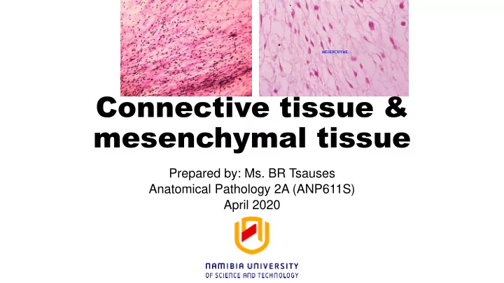
Connective tissue & mesenchymal tissue
Prepared by: Ms. BR Tsauses Anatomical Pathology 2A (ANP611S) April 2020
mesenchymal tissue Prepared by: Ms. BR Tsauses Anatomical Pathology - - PowerPoint PPT Presentation
Connective tissue & mesenchymal tissue Prepared by: Ms. BR Tsauses Anatomical Pathology 2A (ANP611S) April 2020 Learning objectives Understand connective and mesenchymal tissues in order to describe and apply appropriate staining
Prepared by: Ms. BR Tsauses Anatomical Pathology 2A (ANP611S) April 2020