Muscle Tissue Muscle Tissue Gen. Info. Muscle tissue makes up - - PowerPoint PPT Presentation
Muscle Tissue Muscle Tissue Gen. Info. Muscle tissue makes up nearly half the bodys mass, with over 700 different skeletal muscles! The main tissue in the heart and walls of hollow organs The terms sarco and mys come from
Muscle Tissue – Gen. Info.
- Muscle tissue makes up nearly half the body’s
mass, with over 700 different skeletal muscles!
- The main tissue in the heart and walls of
hollow organs
- The terms sarco… and mys… come from greek
words meaning “flesh” & “muscle”.
2
Overview of Muscle Tissue
I. Functions of muscle tissue
1. Movement
A. Skeletal muscle ‐ attached to skeleton creates movements of the body (joints & leverage systems) B. Smooth muscle – squeezes fluids and other substances through hollow organs C. Regulates entrance and exit of materials into, through & out of body
2. Maintenance of posture 3. Joint stabilization 4. Thermogenesis 5. Support & Protection of soft tissues/organs
3
Overview of Muscle Tissue
II. Characteristics of Muscle Tissue
1. Contractility 2. Excitability 3. Extensibility 4. Elasticity
4
III. Types of Muscle Tissue
1. Skeletal muscle tissue – packaged into skeletal muscles (700+)
A. Makes up 40% of body weight
2. Cardiac muscle tissue 3. Smooth muscle tissue – occupies the walls of hollow organs
5
Overview of Muscle Tissue
Muscle Comparison Chart
6
Skeletal Cardiac Smooth Cylindrical Cylindrical & branched Fusiform Yes Yes No Multi- nucleate & peripheral Uninucleate & central Uninucleate & central Voluntary Involuntary Involuntary none Intercalated discs May be single-unit
- r multi-unit
Muscle Tissue Cell Shape Striae Nucleus Control Special structures
Commonalities of Muscle Tissue
- Cells of skeletal and smooth muscles are known as
muscle fibers
- Muscle contraction depends on two types of
myofilaments
– One type contains globular actin and other proteins – Another type contains myosin
- It is the action of myosin on actin that allows these two proteins to
generate contractile force
– Muscle contraction occurs along its longitudinal axis only
- Plasma membrane is called a sarcolemma
- Cytoplasm is called sarcoplasm
7
Skeletal Muscle
- Each muscle is an organ that . . .
– Consists mostly of muscle tissue – but also contains
– Connective tissue – strong attachments continuous through muscle structure – Blood vessels – nourishment & waste removal – Nerves – communication & control
8
Basic Features of a Skeletal Muscle
I. Connective tissue and fascicles
A. Connective tissue sheaths bind a skeletal muscle and its fibers together
1.
Epimysium – dense regular connective tissue surrounding entire muscle
2.
Perimysium – surrounds each fascicle (group of muscle fibers)
3.
Endomysium – a fine sheath of connective tissue wrapping each muscle cell
B. Connective tissue sheaths are continuous with tendons
9
10
Basic Features of a Skeletal Muscle
Muscle fiber endomysium fascicle perimysium muscle bundle epimysium All the fibrous sheaths are continuous to the tendon! - why? Where is the contractile event? Where is the force applied? Line view of the relationship between the connective tissue coverings.
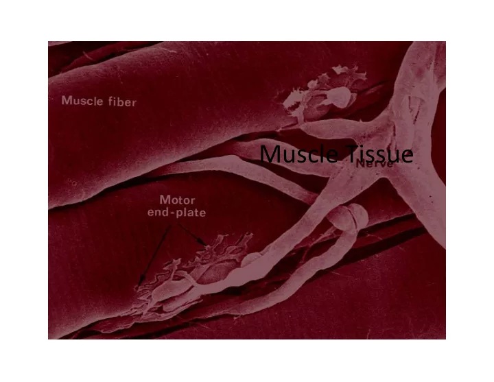
Basic Features of a Skeletal Muscle
II. Innervation & Vasculature
A. Each skeletal muscle has innervations and vasculature which becomes an integral portion of the muscle B. The motor unit consists of the muscle fibers, blood vessels and the nerve that innervates it.
1. Nerves
a. Efferent Motor nerves enter the muscle and divide continually until each motor unit has innervation, the neuromuscular junction. b. Afferent sensory nerves provide feedback about muscle tension
2. Arteries & Veins
a. Arteries enter and divide continually until each muscle fiber has a capillary network around it to provide the muscle with oxygen, nutrients and provide a route for removal of waste (carbon dioxide, heat, lactic acid. . .)
11
Basic Features of a Skeletal Muscle
III. Muscle attachments
A. Most skeletal muscles run from one bone to another,
- r possibly span across
more than one bone (biarticular muscle ex. flexor carpi radialis) B. One bone will move –
- ther bone remains fixed
- r stable
1. Insertion – more movable attachment 2. Origin – less movable attachment
12
Basic Features of a Skeletal Muscle
III. Muscle attachments (continued)
C. Muscles attach to origins and insertions by connective tissue
1. Fleshy attachments – connective tissue fibers are short
1.
- Ex. pectoralis major . . On the origin side
2. Indirect attachments – connective tissue forms a tendon or aponeurosis
1. Visible anytime a longer “cord” is present, or a broad flat sheet.
3. Skin, cartilage, sheets of fascia or raphe (line/ridge of a ligament)
D. Bone markings present where tendons meet bones
1. Tubercles, trochanters, tuberosities, crests… any roughened surface
13
Microscopic and Functional Anatomy
- f Skeletal Muscle Tissue
I. The skeletal muscle fiber
A. Fibers are long and cylindrical
1. Are huge cells – diameter is 10‐100µm 2. Length – several centimeters to dozens of centimeters
B. Each cell formed by fusion of many embryonic cells C. Cells are multinucleate (due to above) D. Nuclei are peripherally located
1. Due to the majority of the cell’s internal space taken up by the myofilaments.
E. Cell membrane is called the sarcolemma
14
Microscopic and Functional Anatomy
- f Skeletal Muscle Tissue
- F. Striations result from internal structure of
myofibrils
- G. Myofibrils – long rods within cytoplasm
- 1. Make up 80% of the cytoplasm
- 2. These contain the smallest contractile units of a
muscle in repeating segments called sarcomeres.
15
Diagram of Part of a Muscle Fiber
16
Figure 10.4b
Important Components We Will Consider
- The neuromuscular junction (NMJ)
- The sarcolemma
- The t‐tubules and sarcoplasmic reticulum (SR)
- The sarcomere it’s filaments
- The structural aspect of muscle contraction
17
The NMJ, Sarcolemma, T Tubules & SR I. What is it’s role in muscle contraction?
A. Action potential is transferred to the sarcolemma at the neuromuscular junction B. Impulse travels along the sarcolemma of the muscle cell (multidirectionally)
1. Impulses further conducted by t tubules a. T tubule – a deep invagination of the sarcolemma b. The close proximity of the t tubules to the sarcoplasmic reticulum causes it (SR) to depolarize, opening Ca gated channels, allowing Ca to diffuse rapidly out of the SR.
18
The NMJ, Sarcolemma, T Tubules & SR
- Sarcoplasmic reticulum is specialized smooth
ER
– Interconnecting tubules surround each myofibril
- Some tubules form cross‐channels called terminal
cisternae
- Cisternae occur in pairs on either side of a t tubule
– Contains calcium ions
- released when muscle is stimulated to contract after the
events at the neuromuscular junction
– Calcium ions diffuse through cytoplasm
- Trigger the sliding filament mechanism
– bind to troponin. . . at the sarcomere level..
19
Sarcomere
- The basic unit of contraction of skeletal
muscle
– Z disc (Z line) – boundaries of each sarcomere – Thin (actin) filaments – extend from Z disc toward the center of the sarcomere – Thick (myosin) filaments – located in the center of the sarcomere
- Overlap inner ends of the thin filaments
- Contain ATPase enzymes
20
Sarcomere Structure
- A bands – full length of the thick filament
– Includes inner end of thin filaments
- H zone – center part of A band where no thin
filaments occur
- M line – in center of H zone
– Contains tiny rods that hold thick filaments together
- I band – region with only thin filaments
– Lies within two adjacent sarcomeres
21
Sarcomere and Myofibrils
22
Sliding Filament Structures
23
Figure 10.6a
Changes in Striation During Contraction
24
Microscopic and Functional Anatomy
- f Skeletal Muscle Tissue
- Muscle extension
– Muscle is stretched by a movement opposite that which contracts it
- Muscle fiber length and force of contraction
– Greatest force produced when a fiber starts out slightly stretched – Myosin heads can pull along the entire length of the thin filaments
25
The Role of Titin
- Titin – a spring‐like molecule in sarcomeres
– Resists overstretching – Holds thick filaments in place – Unfolds when muscle is stretched
26
Neuromuscular Junction
27
Sarcoplasmic Reticulum and T Tubules in the Skeletal Muscle Fiber
28
Sarcoplasmic reticulum network and terminal cisternae
29
Types of Skeletal Muscle Fibers
- Skeletal muscle fibers are categorized
according to:
– How they manufacture energy (ATP) – How quickly they contract
- Skeletal muscle fibers are divided into three
classes:
– Slow oxidative fibers (Type I) – Fast oxidative fibers (Type IIa) – Fast glycolytic fibers (Type IIx) – Fast glycolytic fibers (Type IIb)
30
31
Fiber Type Type I fibers Type II a fibers Type II x fibers Type II b fibers Contraction time Slow Moderately Fast Fast Very fast Size of motor neuron Small Medium Large Very large Resistance to fatigue High Fairly high Intermediate Low Activity Used for Aerobic Long‐term anaerobic Short‐term anaerobic Short‐term anaerobic Maximum duration of use Hours <30 minutes <5 minutes <1 minute Power produced Low Medium High Very high Mitochondrial density High High Medium Low Capillary density High Intermediate Low Low Oxidative capacity High High Intermediate Low Glycolytic capacity Low High High High Major storage fuel Triglycerides Creatine phosphate, glycogen Creatine phosphate, glycogen Creatine phosphate, glycogen
Cardiac Muscle Tissue
I. General Characteristics
A. Occurs exclusively in heart wall B. Forms a thick layer called myocardium C. Striated like skeletal muscle D. Contracts by sliding filament mechanism E. Single cells – not fused colonies like skeletal muscle F. Separated by delicate endomysium G. Cells are branched H. Join at intercalated discs – complex junctions I. Form cellular networks J. Each cell typically contains one centrally‐located nucleus
32
Cardiac Muscle Tissue
33
Cardiac Muscle Tissue
- Not all cardiac cells are innervated
– Innervation comes from the autonomic nervous system for controlling rate/contractility. – Normal contractions are initiated by the sino‐atrial node (SA node) “pacemaker”, causing the heart to be “autorhythmic” – The SA node when activated initiates an electrical conduction system that eventually reaches the myocardium (more when we get to the heart).
34
Cardiac Muscle Tissue
- Contraction is triggered to by Ca2+ entering
the sarcoplasm
– Action potentials from the specialized conduction myofibers (Purkinje fibers) trigger the sarcoplasmic reticulum to release Ca2+ ions – Ions diffuse into sarcomeres
- Triggers sliding filament mechanism
35
Cardiac Muscle Tissue
- Intercalated discs – complex junctions
– Adjacent sarcolemmas interlock – Possess three types of cell junctions
- Desmosomes
- Fasciae adherans – long desmosome‐like junctions
- Gap junctions
36
Cardiac Muscle Cell
37
Smooth Muscle Tissue
- Found in the walls of visceral organs
– Six major locations
- Walls of circulatory vessels
- Respiratory tubes
- Digestive tubes
- Urinary organs
- Reproductive organs
- Inside the eye
38
Smooth Muscle Tissue
- Cells are spindle‐shaped
- Contain one centrally located nucleus
- Separated by endomysium
- Grouped into sheets in walls of hollow organs
– Longitudinal layer – muscle fibers run parallel to
- rgan’s long axis
– Circular layer – muscle fibers run around circumference of the organ
- Both layers participate in peristalsis
39
Smooth Muscle Tissue
- Cross‐section of intestines showing
longitudinal and circular layers
40
Smooth Muscle Tissue
- Cells are non‐striated and contain no
sarcomeres
- Thin and thick filaments are present
- Entry of Ca2+ into the cytoplasm signals
contraction of the smooth muscle fiber
- Caveolae – tiny infoldings of the sarcolemma
41
Smooth Muscle Cells
42
Smooth Muscle Tissue
- Contraction is slow and sustained
– Takes 30 times longer to contract than skeletal muscle fiber – Maintains contractile force for a long time
- Is innervated by autonomic nervous system
– Only a few smooth muscle fibers are innervated in each sheet
- Impulse spreads through gap junctions
- Whole sheet contracts as a unit (single unit smooth muscle)
– Innervations on the individual cells create smooth muscle with more control (multi‐unit smooth muscle).
- iris of the eye and arrector pili muscles
43