muscle muscle muscle muscle cytoskeleton cytoskeleton
play

Muscle Muscle Muscle Muscle Cytoskeleton Cytoskeleton - PDF document

8/27/2018 Muscle Muscle Muscle Muscle Cytoskeleton Cytoskeleton Cytoskeleton Cytoskeleton II II II II Balazs Kiss Postdoc at Granzier Lab Department of Cellular and Molecular Medicine, University of Arizona, Tucson, AZ 85721 Email:


  1. 8/27/2018 Muscle Muscle Muscle Muscle Cytoskeleton Cytoskeleton Cytoskeleton Cytoskeleton II II II II Balazs Kiss Postdoc at Granzier Lab Department of Cellular and Molecular Medicine, University of Arizona, Tucson, AZ 85721 Email: kissb@email.arizona.edu
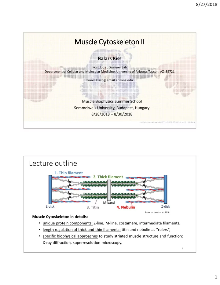
 Muscle Biophysics Summer School Semmelweis University, Budapest, Hungary 8/28/2018 – 8/30/2018 http://www.rsc.org/images/b901714c-400-FOR-TRIDION_tcm18-152053.jpg Lecture outline 1. Thin filament 2. Thick filament M-band Z-disk Z-disk 3. Titin 4. Nebulin based on Labeit et al., 2010. Muscle Cytoskeleton in details: • unique protein components: Z-line, M-line, costamere, intermediate filaments, • length regulation of thick and thin filaments: titin and nebulin as “rulers”, • specific biophysical approaches to study striated muscle structure and function: X-ray diffraction, superresolution microscopy. 2 1

  2. 8/27/2018 Muscle cytoskeleton at the Z-disks Henderson and Gomez et al, 2017. Z-disks: borders of contractile units: • thin filament anchoring: α-actinin , filamin, myotilin, • force transmission: via titin and nebulin, • signaling node: muscle LIM protein (MLP), muscle ankyrin repeat proteins (MARPs), myopodin, myopalladin. 3 α-Actinin • α-actinin 2: cardiac muscle • α-actinin 3: fast skeletal and cardiac muscle • head-to-tail antiparallel homodimers • contour length: 35 nm • N-term: actin binding (ABD) • C-term: 2 EF hand domains: titin binding • 4 spectrin-repeats (SR) • mutations: ACTA2 : DCM, HCM ACTA3 : nonsense - 16% of humans! 4 2

  3. 8/27/2018 α-Actinin at the Z-disks Z-disk I-band I-band Grison et al, 2017. 5 α-Actinin-Titin interaction at the Z-disk Titin at the Z-disk: Testing the mechanical stability: • Z1-Z2 (Ig-domains): binds telethonin • using optical trap • Z-repeats 1-7: could bind α-actinin • dsDNA spacer towards the microbeads • short covalent linker between titin Z- repeat and α-actinin EF domain Joseph et al, 2001 M Z Grison et al, 2017. Luther, 2009 6 3

  4. 8/27/2018 Analysis of kinetic parameters – Hidden Markov Model Life of a Grad Student 3-state model • lab • coffee-shop • bar Analysis of kinetic parameters – 3 state model fixed trap distance (passive mode) A: folded A: folded A: unfolded T: bound T: unbound T: unbound (FB) (FU) (FB) 4

  5. 8/27/2018 α-Actinin-Titin interaction model • different Z-repeats have different affinities to α- actinin, • avidity: accumulated strength of multiple, Grison et al, 2017. parallel bonds/interactions. 9 Filamin-C and Myotilin at Z-disk Filamin-C: • connecting element to integrins, • affinity to its targets might be modulates by mechanical forces , • nonsense mutation: myofibrillar myopathy. Rognoni et al, 2012. Myotilin: • homodimers, • prevents actin depolymerization, • interacts with multiple Z-disk proteins • nonsense mutation: “myotilinopathies”. Sanfilippo and Di Rosa, 2016. 10 5

  6. 8/27/2018 M-band: hub for mechanosensing • M-bridges: stabilize myosin in the M-band center of the sarcomere • Myomesin-1: all striated muscles • Myomesin-2 (M-protein): fast (type IIb/x) muscle fibers • Myomesin-3: intermediate twitch fibers • antiparallel homodimers, dimerization through the C-terminal Ig-domain • domains are able to stretch to 2.5x Gautel and Djinović-Carugo, 2016. their original length: “elastic band” 11 M-band titin: titin kinase unfolding AFM-based molecular combing Mártonfalvi and Kellermayer, 2014. • titin kinase might be unfolded with receding meniscus: mechanosensor 12 6

  7. 8/27/2018 Costamere: protects against mechanical stress Bi-directionally links the cytoskeleton to the extracellular matrix: dystrophin vinculin- glycoprotein talin- complex integrin system Henderson and Gomez et al, 2017. 13 Costamere pathology: myopathies • BMD: Becker muscular dystrophy • DMD: Duchenne muscular dystrophy • CMD1C-1D: congenital muscular dystrophy type 1C-1D • FCMD, Fukuyama congenital muscular dystrophy • LGMD2C-2F: limb-girdle muscular dystrophy type 2C-2F • LAMA2: laminin alpha 2 chain or Henderson et al, 2017. merosin-deficient muscular dystrophy • MEB: muscle-eye-brain disease • WWS: Walker–Warburg syndrome 14 7

  8. 8/27/2018 Intermediate filaments: scaffold for cell organelles • diameter: 8-10 nm • coiled-coil dimer: head tail • main components in muscle: • desmin (2% of total protein in heart) • cytokeratins (K8K19) • desmin knockout mice: myofibril misalignment, loss of nuclear shape, impaired force generation • mutations: “desminopathies” 15 Molecular elasticity of desmin AFM-based Desmin assembly shape fluctuation analysis with Mg 2+ • curve fit: persistence length (P): ~0.45 µm for lateral bending Kiss et al, 2011. 16 8

  9. 8/27/2018 Nebulin in the sarcomere Tmod M-band Z-disk Z-disk Nebulin • ~850 kDa, ~1µm • only in skeletal muscle • 35 aa residue modules ( M ) • super repeat (SR) from 7M • mutations: nemaline myopathy Kontrogianni-Konstantopoulos et al, 2009. 17 Putative role: thin filament stabilizer? Squire et al, 2004. Actin subunit Nebulin Trinick, 1992. Tropomyosin (one strand, simulation) Nebulin interacts stronglywith thin filament: Troponin • 7 actin monomer binding motifs per SR, (projected image) 2.73 nm 38.5 nm • 1 troponin and tropomyosin binding subunit per SR, • nebulin binds along the long pitch helix of the thin filament, • 2 nebulin molecules per thin filament? 18 9

  10. 8/27/2018 How to measure thin filament compliance? Problem #1: nebulin cannot be expressed due to its giant size. Problem #2: isolation of native nebulin is currently not possible. Solution: mechanical effect of nebulin on the thin filament could be assessed by directly measuring thin filament compliance in living muscle. Background: thin filaments of striated muscle are longitudinally extensible! Myosin spacing change (%) Actin spacing change (%) Huxley et al, 1994. Huxley et al, 1994. Brunello et al, 2014. Brunello et al, 2014. 19 X-ray diffraction in muscle research “Then as now, x-ray diffraction remains the only technique to detect nanometer-scale structural changes in real physiological time in living muscle.” Hitchcock-DeGregori and Irving, 2014. Hugh Esmor Huxley Early studies on sarcomere structure and function : based on X-ray diffraction: • inter-thick filament spacing, • sliding filament theory of contraction; moving cross-bridges, • tropomyosin: steric blocking model, • longitudinal extensibility of thin and thick filaments. 20 10

  11. 8/27/2018 X-ray diffraction: (bio)physical basis Bragg’s Law: 2 d sin θ = nλ when λ=constant: d ~ � d: lattice spacing � θ: angle of incidence λ: wavelength of the electromagnetic wave reciprocal space real space 21 X-ray diffraction on fibers X-ray real space reciprocal space Fibrous protein: naturally ordered structure 22 11

  12. 8/27/2018 X-ray diffraction on skeletal muscle meridian P 1/P skeletal muscle (fiber) subunit meridional spacing pattern equator 1/P X-ray P lattice equatorial spacing pattern real space real reciprocal 1 8.7 2 250 space space reciprocal space 23 Structural data from the diffraction image meridian 27.3 Å 28.6 Å Ebashi et al., 1969. Volkmann et al., 2001. 51 Å 27.3 Å = 2.73 nm: 51 Å and 59 Å: 59 Å actin subunit repeat thin filament helix equator 143 Å 5 th order: 1,1 1,0 28.6 Å Needham, 1971. 28.6 Å = 2.86 nm: Milman, 1998. d 10 , d 11 : lattice spacing myosin backbone spacing I 10 , I 11 : cross-bridge meridional pattern equatorial pattern 24 1 8.7 2 250 12

  13. 8/27/2018 Higher order reflections: fine structure of proteins N.b. changes in spacing and intensity! fundamental periodic mass distribution in axial myosin BACKBONE (7.2 nm) periodicity Huxley and Brown, 1967. axial repeat of myosin HEAD (14.3 nm) sensitive to the helix perturbations ~43-nm fundamental axial periodicity rest tetanus Linari et al, 2000. Park-Holohan et al, 2012. 25 Experimental strategy Ctrl and m. soleus Neb cKO • 50-70 days old • Neb cKO: few percent nebulin expression • force- and tension deficit in Neb cKO 26 13

  14. 8/27/2018 What is the mechanical effect of nebulin on the TF? 0.0075%/kPa 0.0027%/kPa Squire et al, 2004. 2.73nm = 27.3Å: Actin Nebulin actin subunit repeat (one strand, simulation) subunit 27Å • thin filaments of Neb cKO are ~3-times more compliant than Ctrl • nebulin stiffens the thin filament via its actin subunits Kiss et al, in press 27 Nebulin increases single thin filament stiffness. Ultrastructure of muscle cross-section (EM) 100 ECM Void Mito Myo 80 **** **** 60 60 30 0 Ctrl Neb cKO Single thin filament stiffness: EM • significantly reduced mitochondrial • Ctrl: 30.3 pN/nm area in Neb cKO • Neb cKO: 10.0 pN/nm • X-ray significantly increased myofibril area in Neb cKO Kiss et al, in press 28 14

Download Presentation
Download Policy: The content available on the website is offered to you 'AS IS' for your personal information and use only. It cannot be commercialized, licensed, or distributed on other websites without prior consent from the author. To download a presentation, simply click this link. If you encounter any difficulties during the download process, it's possible that the publisher has removed the file from their server.

Recommend


More recommend