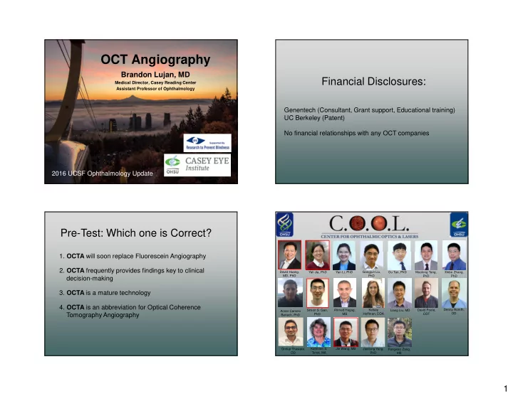

OCT Angiography Brandon Lujan, MD Financial Disclosures: Medical Director, Casey Reading Center Assistant Professor of Ophthalmology Genentech (Consultant, Grant support, Educational training) UC Berkeley (Patent) No financial relationships with any OCT companies 2016 UCSF Ophthalmology Update Pre-Test: Which one is Correct? 1. OCTA will soon replace Fluorescein Angiography 2. OCTA frequently provides findings key to clinical David Huang, Yali Jia, PhD Yan Li, PhD Gangjun Liu, Ou Tan, PhD Maolong Tang, Xinbo Zhang, MD, PhD decision-making PhD PhD PhD 3. OCTA is a mature technology 4. OCTA is an abbreviation for Optical Coherence Kelsey Denny Romfh, Simon S. Gao, Ahmed Hagag, Liang Liu, MD David Poole, Acner Camino Tomography Angiography Hoffman, COA OD PhD MS COT Benech, PhD Jie Wang, MS Omkar Thaware, Kathleen S. Jianlong Yang, Pengxiao Zang, OD Torok, MA PhD MS 1
Retinal Capillary Plexuses Retinal Capillary Plexuses Courtesy of Dave Wilson, MD Casey Eye Institute NFL Nerve Fiber Layer Radial GCL Superficial Ganglion Cell Layer INL Intermediate Inner Nuclear Layer Deep Ern Zi Tan et al, 2012 5 OCTA is Motion Contrast OCTA is Motion Contrast 2
OCTA is Motion Contrast OCTA is Motion Contrast OCTA is Motion Contrast OCTA is Motion Contrast 3
OCTA is Motion Contrast OCTA is Motion Contrast OCTA is Motion Contrast Optical Coherence Tomography Angiography Decorrelation Signals Simple Standard Deviation Map 4
FDA-Approved Commercial OCTA Systems Zeiss Angioplex Optovue Avanti Zeiss Angioplex Optovue Avanti Several more on the way... 45yo CRVO 20/25 5
Baseline Inject 20/25 +6W Observe 20/20 20/25 +12W Inject +18W Inject 20/25 +24W Observe 20/16* Inner retinal atrophy +24W: Temporal ischemia with new scotoma Inner and middle retinal ischemia / atrophy +18W +24W 6
OCTA OCTA Angiography Fluorescein Angiography +18W +24W Many capillaries better visualized on OCTA ...but not with slow flow! Depth Encoding MacTel Type 2 Phase-Variance OCT Angiography Fluorescein Angiogram 00:37 Zeiss AngioPlex Schwartz DM et al, 2014 Courtesy of Philip Rosenfeld, MD, PhD 7
Projection Artifacts in OCTA B-scans Projection Artifacts in OCTA Slabs Consequence of decorrelation "shadows" Consequence of decorrelation "shadows" Superficial plexus Intermediate plexus Decorrelation: Real Graph-based segmentation Real Projection Vessels artifacts Vitreous-ILM Above IPL-INL Below IPL-INL Below sOPL Projection Projection Decorrelation: Artifact RPE-BM artifacts artifacts Structural OCT Inner retinal flow Outer retinal flow Choroidal flow Deep plexus Outer retina Normal Retinal Anatomy Zhang M, et al. 2016 Projection suppression by amplitude Projection suppression by simple filtering: Projection-Resolved OCTA Slab Subtraction Superficial plexus Intermediate plexus Superficial plexus Intermediate plexus Real Projection Vessels artifacts Fragment Real Projection Graph-based segmentation Graph-based segmentation Vessels artifacts Vitreous-ILM Vitreous-ILM Above IPL-INL Above IPL-INL Fragmented Below IPL-INL Below IPL-INL OK Below sOPL Below sOPL Projection Projection RPE-BM RPE-BM artifacts artifacts Projection Structural OCT Projection Structural OCT Inner retinal flow artifacts Inner retinal flow artifacts Outer retinal flow Outer retinal flow Choroidal flow Choroidal flow Deep plexus Outer retina Deep plexus Outer retina Normal Retinal Anatomy Normal Retinal Anatomy Zhang M, et al. 2016 Zhang M, et al. 2016 8
PR-OCTA Details Retinal Vascular Anatomy Macular Ischemia in Non-Proliferative DR Superficial Intermediate Deep Inner Case 1 C D A B Case 2 C D A B 3 retinal plexuses show different patterns of vascular pathologies Peripapillary Parafoveal Peripheral Zhang M et al, 2016 Rapid Dynamic CNV Response to Anti-VEGF Post-Test: Which one is Correct? Inner retinal flow Outer retinal flow (CNV) Anti-VEGF 1. OCTA complements Fluorescein Angiography Baseline 2 days 1 week 4 weeks 2. OCTA provides insights into disease pathogenesis 1 st Rx 3. OCTA is a rapidly developing technology 1 day 2 weeks 6 weeks 4. OCTA is an abbreviation for Optical Coherence Tomography Angiography 2 nd Rx 5. All of the Above Max response Huang D et al, 2015 9
Save the date! OCT A NGIOGRAPHY S UMMIT July 21 - 22, 2017 Portland, Oregon, USA 10
Recommend
More recommend