
SLIDE 1
2016
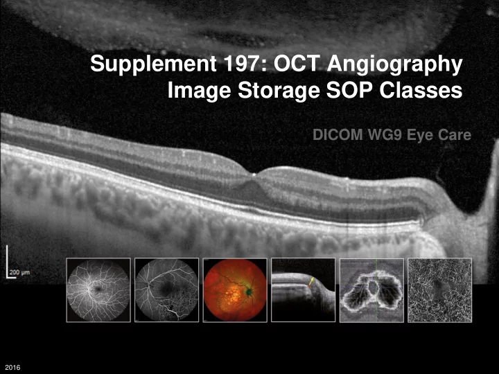
Supplement 197: OCT Angiography Image Storage SOP Classes DICOM WG9 - - PowerPoint PPT Presentation
Supplement 197: OCT Angiography Image Storage SOP Classes DICOM WG9 Eye Care 2016 OCT-A Separating Vascular Networks 2016 OCT-Angiography - Outcome Clinicians typically make their assessment based on the OCT En face images derived from OCT
2016
2016
2016
2016
2016
Patient with mCNV. FA/ICGA images (top) show capillary leakage; OCT-A (bottom) provides a superior visualization of the newly formed vessels and depth-resolved resolution of vascular perfusion – neovascularization is clearly visible in deeper layers (right) than in superficial layers (left).
2016
2016
2016
2016
2016
2016
2016
2016
2016
2016
2016
2016
2016
2016
2016
2016
2016
2016