SLIDE 1
5/26/2016 1 Cribriform Lesions of the Breast
Yunn-Yi Chen, MD, PhD Professor Director of Immunohistochemistry Laboratory UCSF
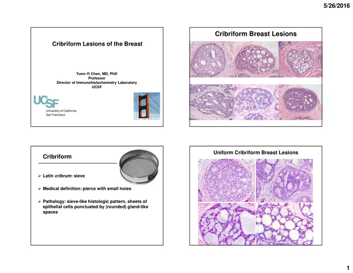
Cribriform Breast Lesions
Cribriform
Latin cribrum: sieve Medical definition: pierce with small holes Pathology: sieve-like histologic pattern, sheets of epithelial cells punctuated by (rounded) gland-like spaces
Uniform Cribriform Breast Lesions
SLIDE 2 5/26/2016 2
Cribriform DCIS Invasive Cribriform Carcinoma Adenoid Cystic Carcinoma Collagenous Spherulosis
Cribriform Breast Lesions
Lobulocentric vs diffuse pattern Single vs dual cell populations Luminal contents IHC and molecular markers Myoepithelial cell (MEC) markers LMW CK, CK5/6 ER Other stains: CD117 (C-Kit), S100 FISH
Approach for Cribriform Lesions Cribriform DCIS-- Lobulocentric
Diagnostic accuracy of DCIS by pathologists
(Results of ECOG Trial 5194)
49/693 (7.1%): misclassified
(Simpson J, et al: USCAP meeting abstract, 2011, 64A)
ADH 28 (57%) UDH 5 (10%)
LCIS 5 Papilloma 2 Radial scar 2 Invasive carcinoma 3 Columnar cell lesion 2 Papillary apocrine change 1 Mucocele-like lesion 1
SLIDE 3 5/26/2016 3
Distinction from ADH and UDH Criteria based on cytology, architecture and extent
- Cytology-- monotonous, round to oval nuclei, distinct
cell membrane
- Architecture-- rounder/rigid, polarized to lumen
- Extent-- complete involvement of spaces, continuous
span > 2 mm (3 mm by some authors and arising in papilloma)
Inter-observer variability
- Share with your colleagues
Adjunct: HMWK (CK5/6), ER
- UDH-- CK5/6 positive (mosaic pattern), variable ER
- ADH and DCIS– CK5/6 negative, diffuse and strong ER
Diagnosis of low grade Cribriform DCIS Cribriform DCIS UDH
Monotonous Evenly spaced Distinct cell membrane Nuclei polarized to lumen Heterogeneous Crowded and overlapping Indistinct cell membrane Nuclei lack of polarization
Cribriform DCIS UDH
Nuclei perpendicular to lumen Nuclei parallel to lumen
SLIDE 4
5/26/2016 4 Usual Ductal Hyperplasia
CK5/6 ER
Atypical Ductal Hyperplasia--
Combined ER (brown) and CK5/6 (red) stain Negative CK5/6, diffuse and strong ER
Necrosis may be seen in UDH
SLIDE 5 5/26/2016 5 UDH with necrosis
CK5/6 ER
Extent criteria for low-grade DCIS--
complete involvement of > 2 mm
2 mm
ADH ADH LG DCIS
CK5/6, p63 (brown)/LMW CK (red)
Benign Apocrine Proliferation
- Apocrine cells are CK5/6 and ER negative
- Uniform nuclei, fine chromatin
- No necrosis
DCIS with apocrine features
- Significant (≥3x) variation in nuclear size
- Irregular nuclear membrane, coarse chromatin
- Often with comedo necrosis
SLIDE 6 5/26/2016 6
Cribriform DCIS Involving a Radial Sclerosing Lesion (RSL)
Cribriform DCIS in RSL-- Mimic invasion Reduced MEC staining p63 SMM
(Hilson et al: Am J Surg Pathol 2010;34:896-900)
Lobulocentric Single luminal type epithelial cells Luminal contents: calcifications Immunophenotype MEC around the duct space ER: +++ CAM5.2 +++, CK5/6 -
Cribriform DCIS
Benign Aggregates of eosinophilic fibrillar spherules or myxoid material (BM material) Biphasic myoepithelial and epithelial proliferation Two types of lumens
Collagenous Spherulosis
SLIDE 7
5/26/2016 7
Collagenous Spherulosis: Lobulocentric
Collagenous Spherulosis Arising in a Papilloma
SLIDE 8
5/26/2016 8
Coll IV SMM p63 Collagenous Spherulosis-- two cell populations
Myoepithelial cells pseudolumen Epithelial cells true glandular space
CAM5.2 SMM
Coll Spherulosis Cribriform DCIS
Collagenous Spherulosis-- Core Biopsy for Calcifications
SLIDE 9
5/26/2016 9
Collagenous Spherulosis in Radial Scar
Monomorphic cells with cribriform architecture ? DCIS
SLIDE 10
5/26/2016 10
LCIS involving collagenous spherulosis-- Pitfall in mimicking cribriform DCIS
E-cad E-cad p63
LCIS involving collagenous spherulosis-- positive E-cad staining from ME cells Lobulocentric Dual MEC and epithelial cells MEC: pseudolumens, BM material Epithelial cells: true glandular spaces, ± eosinophilic secretion Immunophenotype MEC markers + around pseudolumens LMW CK (CAM5.2) + around true glandular spaces
Collagenous Spherulosis
Invasive cribriform CA
Adenoid cystic CA
Invasive Carcinoma with Cribriform Pattern
SLIDE 11
5/26/2016 11
Invasive Cribriform Carcinoma
Excellent prognosis Luminal A molecular type Cribriform pattern invading stroma Often mixed with other invasive (tubular carcinoma in 17-23%) Diagnosis--
> 90% cribriform pattern or >50% cribriform + tubular as the second component
Invasive Cribriform Carcinoma-- Diffuse
Inv cribriform CA-- irregular contours, desmoplastic stroma
Cribriform DCIS: Lobulocentric
SLIDE 12 5/26/2016 12
DCIS: Smooth Contours, Myoepithelial Cells
Cribriform DCIS Inv Cribriform CA
Irregular contour Desmoplastic stroma
Inv cribriform CA-- smooth contours, mimic cribriform DCIS
p63
SLIDE 13
5/26/2016 13
Smooth ME-like cells Invasive Irregular In situ p63 p63 SMM
Invasive and in situ cribriform carcinoma
Diffuse pattern Single luminal type epithelial cells Luminal contents: -/+ calcifications, mucin Immunophenotype MEC markers: - ER: +++ CAM5.2 +++; CK5/6 -
Invasive Cribriform Carcinoma
Invasive Cribriform CA Cribriform DCIS
Infiltrate between normal lobules and ducts Normal ductal and lobular architecture preserved Irregular, sharp, angulated edges Smooth, rounded contours Desmoplastic Normal stroma No myoepithelial cells Intact myoepithelial cells
SLIDE 14 5/26/2016 14
Histology similar to salivary gland counterpart Various architectural patterns
- Cribriform
- Tubular/trabecular, solid/basaloid
Triple negative, basal-like molecular type
- Lower mutation rate compared to TN IDC
t(6;9) MYB-NFIB translocation Favorable prognosis
Adenoid Cystic Carcinoma Adenoid Cystic Carcinoma-- Diffuse
Adenoid Cystic CA
Adenoid Cystic CA
* * *
pseudolumen true lumen
Adenoid Cystic Carcinoma
SLIDE 15
5/26/2016 15
* * * *
Adenoid Cystic Carcinoma Adenoid cystic carcinoma Epithelial-like Myoepithelial-like
Immunophenotype
biphasic differentiation
May demonstrate an “aberrant/variable” phenotype
Adenoid Cystic Carcinoma
p63 SMM CK7
Adenoid Cystic CA
SLIDE 16 5/26/2016 16
Adenoid cystic carcinoma-- dual cell populations
Myoepithelial-like/basaloid cells: p63 +, calponin – (brown) Epithelial cells: CAM5.2 + (red)
breast triple stain
Diffuse pattern Dual cell types-- Myoepithelial-like/basaloid cells: pseudolumens with BM material Epithelial cells: true glandular spaces ± secretion Immunophenotype MEC markers: variable; usually p63 +, SMA +/- & SMM/calponin -/+ in basaloid cells LMW CK (CK7) + in epithelial cells ER/PR/HER2 -; CD117 & CK5/6 + (basal-like) MYB +
Adenoid Cystic Carcinoma
Adenoid Cystic Carcinoma
CK5/6 CD117 MYB
Adenoid Cystic Carcinoma--
Excellent Prognosis on population-based studies
Study Population Year # patients LN met Survival*
Ghabach
Surveillance, Epidemiology and End Results Program (SEER)
1977- 2006 338 14/335 (4.2%) 5 y RS: 98.1% 10 y RS: 94.9% 15 y RS: 91.4% Thompson
California Cancer Registry (CCR)
1988- 2006 244 12/244 (4.9%) 5 y RS: 95.6% 10 y RS: 94.9% Kulkarni
National Cancer Data Base
1998- 2008 933 36/703 (5.1%) 5 y OS: 88% (*RS: relative survival; OS: overall survival)
(Ghabach: Breast Cancer Res 2010; Thompson: Breast J 2011; Kulkarni: Ann Surg Oncol 2013)
SLIDE 17 5/26/2016 17 56 y F with a breast lesion 56 y F with a breast lesion 56 y F with a breast lesion
Rare histologic subtype of breast cancer: 0.02% Originally as “Juvenile secretory carcinoma” in 1966
- Most common childhood breast ca
Renamed “secretory carcinoma” in 1980s
- ~2/3 pts > 50 y, mean age 53-56 (range 3 to 89)
Various growth patterns
- Microcystic/cribriform
- Papillary, solid, tubular
Secretory Carcinoma
SLIDE 18 5/26/2016 18
Triple negative or low hormonal receptor expression, basal-like molecular type t(12;15) ETV6-NTRK3 translocation Favorable prognosis
- Lower mutation rate than TN IDC
- Simplex genomic profile by chromosomal analysis
Secretory Carcinoma Secretory Carcinoma-- Diffuse Secretory Carcinoma--
Abundant eosinophilic to amphophilic secretion
Secretory Carcinoma--
- Abundant eosinophilic vacuolated cytoplasm
- Medium-sized oval nuclei, small nucleoli; low mitotic activity
- SBR grade 1 and 2
SLIDE 19 5/26/2016 19 Secretory Carcinoma
PASD stain Secretory material: PASD and Alcian blue + S100 and mammaglobin +++ ER/PR/HER2 - or low ER/PR CK5/6 +
Secretory Carcinoma Secretory Carcinoma
S100 Stain Mammaglobin Stain
Secretory Carcinoma
ER CK5/6
- ER/PR/HER2 – or low ER/PR
- CK5/6 +
- Basal-like
SLIDE 20 5/26/2016 20 Secretory Carcinoma
- Most microcystic/cribriform
glands are invasive
- In situ component may be present
SMM SMM
Secretory Carcinoma--
t(12;15) with ETV6-NTRK3 gene fusion detected by ETV6 split apart FISH probe
ETV6 NTRK6
T(12;15)
Mammary secretory carcinoma Infantile fibrosarcoma Cellular congenital mesoblastic nephroma Some myeloid acute leukemia Mammary analogue secretory carcinoma (MASC) of the salivary gland Cutaneous MASC Some papillary thyroid carcinoma (?MASC of thyroid)
- ~15% of PTC after radiation exposure, 1-4% of sporadic PTC
- One case: TTF1/thyroglobulin - & mammaglobin/S100/GATA3 +
Tumors with ETV6-NTRK3 Translocation
Diffuse pattern Single cell type Extra- and intracellular secretory materials Immunophenotype MEC markers: - in invasive & + in in-situ components S100 and mammaglobin +++ ER/PR/HER2 - or low ER/PR; CK5/6 + (basal-like) t(12;15) with ETV6-NTRK3 fusion gene
Secretory Carcinoma
SLIDE 21 5/26/2016 21
Secretory Carcinoma--
Favorable prognosis on population-based studies
Study Population Year # patients LN met Survival*
Horowitz
Surveillance, Epidemiology and End Results Program (SEER)
1983- 2007 83 29/83 (34.9%) 5 y CSS: 94.4% 10 y CSS: 91.4% (1 pt died of ca) Jacob
National Cancer Database
1998- 2011 246 66/206 (32.0%) OS better than IDC, median not reached (vs 14.8 years for IDC) (*CCS: cause-specific survival; OS: overall survival)
(Horowitz et al: The Breast 2012; Jacob et al: J Surg Oncol 2016)
Cribriform Lesions of the Breast
Yes No
? Pseudolumens with BM material, lined by p63/SMA + cells
Yes No ? Lobulocentric
Coll Spherulosis Cribriform DCIS Adenoid Cystic CA Inv Cribriform CA
SMM + SMM - MEC + MEC -
UDH and ADH Secretory CA
Cribriform Lesions: IHC
Collagenous Spherulosis Adenoid Cystic Invasive Cribriform Cribriform DCIS
SMM
Pos Neg Neg Pos
Calp
Pos Neg Neg Pos
p63
Pos Pos Neg Pos
SMA
Pos Pos Neg Pos
ER
Pos Neg Pos Pos
Crisi Am J Clin Pathol 2005;124:733 <> Mastropasqua Mod Pathol 2005;18:1277 Azoulay Mod Pathol 2005;18:1623 <> Rabban Mod Pathol 2006;19:1351 Foschini Semin Diagn Pathol 2010;27:77
Cribriform Breast Lesions
Inv cribriform CA Cribriform DCIS Adenoid cystic CA Coll spherulosis
p63 SMM
SLIDE 22
5/26/2016 22
Cribriform Breast Lesions
Cribriform DCIS (> 2mm) LCIS in Coll. Spherulosis Secretory CA Inv Cribriform CA Adenoid Cystic CA UDH
Thank you!