
SLIDE 1
5/23/2015 1
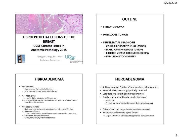
FIBROEPITHELIAL LESIONS OF THE BREAST
UCSF Current Issues in Anatomic Pathology 2015
Gregor Krings, MD PhD Assistant Professor
OUTLINE
- FIBROADENOMA
- PHYLLODES TUMOR
- DIFFERENTIAL DIAGNOSIS
– CELLULAR FIBROEPITHELIAL LESIONS – MALIGNANT PHYLLODES TUMORS – EXCISION VERSUS CORE NEEDLE BIOPSY – IMMUNOHISTOCHEMISTRY
FIBROADENOMA
- Very common
– Most common fibroepithelial lesions – Most common benign tumors of the breast
- Broad age group
– Incidence highest in women <30 years old – Can occur at any age (18.5% of women >40 years old in Breast Cancer Surveillance Consortium)
- Predisposing factors
– No known inherited genetic alterations but risk in some families – Hormonal influence
- Rare in men but associated with gynecomastia, exogenous hormones, drugs
– Cyclosporin A (organ transplant) – Carney complex (myxoid fibroadenomas)
FIBROADENOMA
- Solitary, mobile, “rubbery” and painless palpable mass
- Non-palpable, mammographically detected
- Calcifications (hyalinized fibroadenomas)
- Rarely pain and/or bloody nipple discharge
– Infarction – Pregnancy, prior aspiration procedure, spontaneous
- Often <3 cm but larger tumors not uncommon
- ‘Giant fibroadenomas’ up to 20 cm
– Larger tumors in adolescents (juvenile fibroadenoma)