Disclosures The Approach to the Submandibular Duct and Gland Paid - - PowerPoint PPT Presentation

disclosures
SMART_READER_LITE
LIVE PREVIEW

Disclosures The Approach to the Submandibular Duct and Gland Paid - - PowerPoint PPT Presentation

11/6/2014 Disclosures The Approach to the Submandibular Duct and Gland Paid consultant & Research Support on sleep apnea devices M. Boyd Gillespie, M.D., M.Sc. (Inspire Medical; Olympus; Surgical Specialties) Director, MUSC Salivary


slide-1
SLIDE 1

11/6/2014 1

The Approach to the Submandibular Duct and Gland

  • M. Boyd Gillespie, M.D., M.Sc.

Director, MUSC Salivary Clinic UCSF Salivary Endoscopy Course November 6th, 2014

Disclosures

Paid consultant & Research Support on sleep apnea devices (Inspire Medical; Olympus; Surgical Specialties) Paid consultant on head and neck surgical devices (Medtronic) Izaak Walton The Compleat Angler (1653)

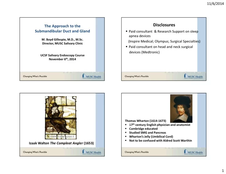
Thomas Wharton (1614-1673)

  • 17th century English physician and anatomist
  • Cambridge educated
  • Studied SMG and Pancreas
  • Wharton’s Jelly (Umbilical Cord)
  • Not to be confused with Aldred Scott Warthin
slide-2
SLIDE 2

11/6/2014 2

North American Experience

  • Ambulatory Operative

Procedure

  • General Anesthesia
  • Nasal Intubation (SMG)
  • Oral Intubation (Parotid)

“Smiley” Cheek Retractor

7

What is the best Endoscope to start with ?

Wharton’s Duct (fully dilated) 2.0-2.5 mm Stenson’s Duct (fully dilated ) 1.5-2.0 mm

cannula: semirigid

  • uter diameter

(0.8) -1.1- (1.6 mm)

We use this size only in about 80% of all patients

Protect Your Scopes

slide-3
SLIDE 3

11/6/2014 3

Methylene Blue

Schaitkin and Marchal Dilators

Wharton’s Duct

Not a straight structure Dips Up Then Down

Dilation

slide-4
SLIDE 4

11/6/2014 4

Kolenda System Kolenda System

Don’t forget Bartholin’s Duct

slide-5
SLIDE 5

11/6/2014 5

Insertion within Wharton´s Duct might be difficult – find duct and open it

Be aware – not in Stensen´s duct (danger of stenosis) Courtesy of Johannes Zenk, MD, PhD

Zeltser, et al. Operative Techniques in Otolaryngology- Head and Neck Surgery 1996; 7: 370-373.

Ductal Scissors

Ductal stenosis > 50% (0.8 mm) Stones > 5mm Ductal anomalies Perforations (saline in FOM) Think Combined Approach

19

slide-6
SLIDE 6

11/6/2014 6

Vessel Loop

slide-7
SLIDE 7

11/6/2014 7

Courtesy of Johannes Zenk, MD, PhD Courtesy of Johannes Zenk, MD, PhD Submandibular Gland Excision Reserved for cases not amendable to minimally invasive approach: Approximately 5% of cases SMG- Usually reserved for fixed, intraglandular submandibular stones below mylohyoid line; Can be performed immediately due to low morbidity. Gland Excision 8 mm fixed, intraglandular left SM stone below Mylohyoid Line

slide-8
SLIDE 8

11/6/2014 8

Gland Excision Intraglandular stone with stenosis

Submandibular Gland Excision Incise low in neck (at least 3 fingers)

Avoid sub-platysmal dissection Deep dissection through soft tissue to posterior digastric Ligation of facial vein (Hayes Martin maneuver) Incise glandular fascia adjacent to digastric Progressively free gland in subfacial plane Ligate facial artery or branches Retract mylohyoid Ligate submandibular ganglion Palpate duct to ensure removal of all stones.

Gland-Preserving Submandibular Surgery Helpful Hints Use Methylene Blue Beginners should try Kolinda Introducer Set Use Tapered Schaitkin Dilators Gently straighten duct with forceps; tease the dilator up then down Passing the diagnostic scope will allow the saline to enlarge the duct making it easier to find Don’t be afraid to make an incision Extensive work on Wharton’s duct may require sublingual gland excision Vessel loops are helpful to retract the duct.