
SLIDE 1
1
Practical Approach to Papillary Breast Lesions
Yunn-Yi Chen, MD, PhD Professor Director of Immunohistochemistry Laboratory Director of Breast Pathology Services UCSF
Overview of papillary lesions Benign intraductal papilloma vs papillary carcinoma Intraductal papilloma with atypia (ADH vs DCIS) Encapsulated (intracystic) papillary carcinoma Solid papillary carcinoma
Outline of Talk
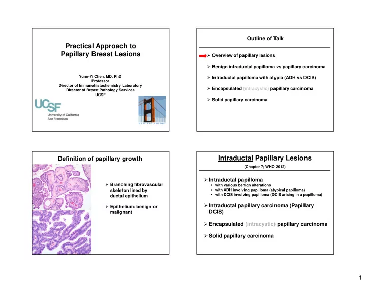
Definition of papillary growth
Branching fibrovascular skeleton lined by ductal epithelium Epithelium: benign or malignant
Intraductal Papillary Lesions
(Chapter 7; WHO 2012)
Intraductal papilloma
with various benign alterations with ADH involving papilloma (atypical papilloma) with DCIS involving papilloma (DCIS arising in a papilloma)