crash course
play

Crash Course Sanofi, Investigator initiated grant A n t h o n y - PowerPoint PPT Presentation

Disclosures Common Orthopaedic and Sports Medicine Problems Founder, RunSafe Founder & CEO, SportZPeak Inc. Crash Course Sanofi, Investigator initiated grant A n t h o n y L u k e MD, MPH, CAQ (Sport Med) University of


  1. Disclosures Common Orthopaedic and Sports Medicine Problems • Founder, RunSafe™ • Founder & CEO, SportZPeak Inc. Crash Course • Sanofi, Investigator initiated grant A n t h o n y L u k e MD, MPH, CAQ (Sport Med) University of California, San Francisco FP Board Review 2015 Overview History is Key Who? • Quick approach to What? MSK problems Pain • Highlight common presentations Instability Dysfunction • Joint by joint • Discuss basics of conservative and surgical management
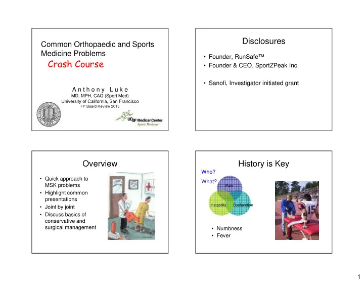
 • Numbness • Fever 1

  2. History is Key Red Flag Symptoms When? • Severe disability • Acute vs Chronic (2 weeks? 6 weeks?) • Numbness and tingling Where? • Night pain • Think anatomy • Constitutional symptoms (fever, wt loss) • One finger test • Swelling with no injury How? • Systemic illness • Mechanism of injury • Multiple joint injury Treatment Options Intrinsic Risk Factors Extrinsic Risk Factors Surgery Conservative • Growth • Training • Reconstruction • MICE (Modified activity, • Anatomy • Technique • Repair Ice, Compression, • Muscle/Tendon • Footwear Elevation) • Re-align imbalance • Surface • Remove internal • Medications/Analgesia derangement • Illness • Occupation • Rehabilitation therapy • Nutrition • Casting/ Braces / • Conditioning • TO PREVENT Orthoses • Psychology INJURIES!! • Crutches 2

  3. Ankle Sprains Physical Exam Mechanism LOOK Symptoms • Inversion, • Swelling/bruising • Localized pain usually plantarflexion (most laterally over the lateral aspect common injury) Anterior FEEL of the ankle talofibular • Eversion (Pronation) • Point of maximal ligament • Difficulty weight tenderness usually bearing, limping ATF Calcaneo • May feel unstable in fibular MOVE ligament the ankle • Limited motion due to swelling Special Tests Anterior Drawer Subtalar Tilt Test Test • Normal ~ 3 mm • Foot in neutral position • Foot in neutral position • Fix tibia • Fix tibia • Invert or tilt calcaneus • Draw calcaneus forward • Tests Calcaneofibular • Tests ATF ligament ligament Sens = 80% Spec = 74% No Sens / Spec PPV = 91% Data NPV = 52% van Dijk et al. J Bone Joint Surg-Br, 1996; 78B: 958-962 3

  4. Subtalar Tilt test Grading Ankle Sprains Grade Drawer/Tilt Pathology Functional Recovery Test results in weeks 1 Drawer and Mild stretch 2 – 4 tilt negative, with no but tender instability 2 Drawer lax, ATFL torn, CFL 4 – 6 tilt with good and PTFL end point intact 3 Drawer and ATFL and CFL 6 – 12 tilt lax injured/torn Ottawa Ankle Rules Treatment of Ankle Sprains • Inability to weight bear immediately and in the Acute Physical Therapy emergency / office (4 steps) • Rest or modified • ROM activities • Strengthening • Bone tenderness at the posterior edge of the • Ice, Compression, • Stretching medial or lateral malleolus (Obtain Ankle Series) Elevation • Proprioception / • Bone tenderness over the navicular or base of • Crutches PRN Balance exercises the fifth metatarsal (Obtain Foot Series) • Bracing (Grade 2 and (i.e. Wobble Board) 3) • Sens 97%, Spec 31-63%, NPV 99%, PPV <20% • Early Motion is essential (Am J Emerg Med 1998; 16: 564-67) 4

  5. Not Always Only a “ Sprain ” “ High Ankle ” Sprains Bone Mechanism Ligaments • Osteochondral talus • Subtalar joint sprain • Dorsiflexion, eversion injury injury • Sinus tarsi syndrome • Lateral talar process fracture • Syndesmotic sprain • Disruption of the • Posterior impingement Syndesmotic ligaments, • Deltoid sprain (os trigonum) most commonly the • Lisfranc injury • Fracture at the base of anterior tibiofibular the fifth metatarsal Tendons ligament • Jones fracture • Posterior tibial tendon • Salter fracture (fibula) • R/O Proximal fibular strain • Ankle fractures fracture • Peroneal tendon subluxation External Rotation Stress Test Squeeze test • Fix tibia • Hold leg at mid calf level • Foot in neutral • Squeeze tibia and • Dorsiflex and fibula together externally rotate ankle • Pain located over anterior tibiofibular ligament area No Sens/ Spec Data Kappa = 0.75 Alonso et al. J Orthop Sports Phys Ther, 1998; 27: 276-284 5

  6. Ankle Sprain Prevention Treatment for Syndesmosis Injury • Ankle braces, tape and Conservative Surgery proprioceptive training help reduce the risk of • Cast or walking boot • May needs ORIF if lateral ankle sprains unstable • Protected Verhagen EALM, van Mechelen W, de Vente weightbearing with W. Clin J Sport Med, 2000 crutches must be painfree • Significant reduction in the number of ankle • PT sprains in people allocated to an external ankle support (RR 0.53, 95% CI 0.40 to 0.69). Handoll et al. Cochrane Database Rev, 2005 Maisonneuve Fracture Acute Hemarthrosis Emergencies 1) ACL (almost 50% in children, >70% in 1. Neurovascular injury adults) 2. Knee Dislocation 2) Fracture (Patella, tibial plateau, Femoral – Associated with multiple ligament injuries supracondylar, Physeal) (posterolateral) 3) Patellar dislocation – High risk of popliteal artery injury – Needs arteriogram • Unlikely meniscal lesions 3. Fractures (open, unstable) 4. Septic Arthritis 6

  7. Anterior Cruciate Ligament (ACL) Urgent Orthopedic Referral Tear • Fracture Mechanism • “ Locked Joint ” - unable to fully extend the • Landing from a • Patellar Dislocation jump, pivoting or decelerating knee (OCD or Meniscal tear) suddenly • Tumor • Foot fixed, valgus stress Anterior Cruciate Ligament (ACL) ACL physical exam Tear LOOK • Effusion (if acute) Mechanism • Landing from a jump, FEEL pivoting or decelerating • “O’Donaghue’s Unhappy Triad” suddenly = Medial meniscus tear, MCL injury, ACL tear • Foot fixed, valgus stress • Lateral meniscus tears more Symptoms common than medial • Audible pop heard or felt • Lateral joint line tender - femoral condyle bone bruise • Pain and tense swelling in minutes after injury MOVE • Feels unstable (bones Double fist sign • Maybe limited due to effusion shifting or giving way) or other internal derangement 7

  8. Special Tests ACL X-ray 20 ° • Lachman's test – test at • Usually non- diagnostic Sens 81.8%, Spec 96.8% 90 ° • Anterior drawer – test at • Can help rule in or Sens 22 - 41%, Spec 97%* out injuries • Pivot shift Sens 35 - 98.4%*, Spec 98%* • Segond fracture – Malanga GA, Nadler SF. avulsion over Musculoskeletal Physical Examination, Mosby, 2006 lateral tibial plateau * - denotes under anesthesia MRI MRI • Sens 94%, Spec 84% • Sens 94%, Spec 84% for ACL tear for ACL tear ACL tear signs ACL tear signs • Fibers not seen in • Lateral femoral corner continuity bone bruise on T2 • Edema on T2 films • May have meniscal tear (Lateral > medial) • PCL – kinked or Question mark sign 8

  9. Initial Treatment ACL Tear Treatment Conservative Surgery • Referral to Orthopaedics/Sports Medicine • No reconstruction • Reconstruction • Consider bracing, crutches • Physical therapy • Depends on activity • Begin early Physical Therapy demands • Hamstring strengthening • Reconstruction allows • Analgesia usually NSAIDs • Proprioceptive training better return to sports • ACL bracing • Reduce chance of symptomatic meniscal controversial tear ADL ’ s • Patient should be • Less giving way symptoms asymptomatic with • Recovery ~ 6 months Special Tests: Meniscus Meniscus Tear Fowler PJ, Lubliner JA. Arthroscopy 1989; 5(3): 184-186. Test Sensitivity Specificity Mechanism Symptoms • Occurs after twisting • Catching Joint line tender 85.5% 29.4% injury or deep squat • Medial or lateral knee Hyperflexion 50% 68.2% • Patient may not recall pain specific injury Extension block 84.7% 43.75% • Usually posterior aspects of joint line McMurray Classic 28.75% 95.3% (Med Thud) • Swelling McMurray Classic (Lat 50% 29% pain) Appley (Comp/Dist) 16% / 5% 9

  10. Modified McMurray Testing X-ray • Flex hip to 90 degrees • May show joint space • Flex knee narrowing and early osteoarthritis changes • Internally or externally rotate lower leg with rotation of knee • Rule out loose bodies • Fully flex the knee with rotations Courtesy of Keegan Duchicella MD MRI Meniscal Tear Treatment Conservative Surgery • Often if degenerative • MRI for specific exam • Operate if internal tear in older patient derangement • Similar treatment to symptoms • Look for fluid (linear mild knee • Meniscal repair if bright signal on T2) osteoarthritis possible into the meniscus • Analgesia • Physical therapy • General Leg Strengthening 10

  11. Medial Collateral Ligament (MCL) Medial Collateral Ligament (MCL) Injury Injury Mechanism Symptoms Physical Exam • Valgus stress to • Pain medially • Tender medially over partially flexed knee MCL (often • May feel unstable proximally) • Blow to lateral leg with valgus “ pseudolocking ” • May lack ROM • Valgus stress test MRI MCL Treatment Conservative Surgery • X-ray non-diagnostic • Analgesia • Rarely needs surgery (rarely avulsion) • Protected motion • MRI not usually +/- hinged brace necessary +/- crutches • Rule out meniscal • Early physical therapy tear 11

Download Presentation
Download Policy: The content available on the website is offered to you 'AS IS' for your personal information and use only. It cannot be commercialized, licensed, or distributed on other websites without prior consent from the author. To download a presentation, simply click this link. If you encounter any difficulties during the download process, it's possible that the publisher has removed the file from their server.

Recommend


More recommend