
SLIDE 1
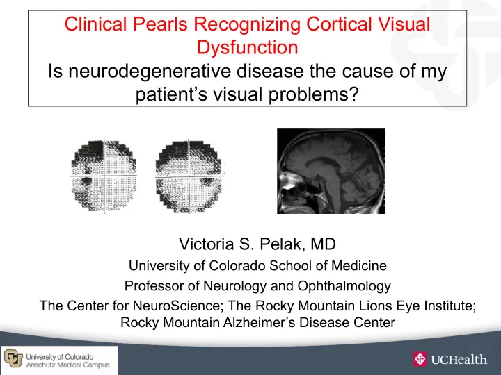
Clinical Pearls Recognizing Cortical Visual Dysfunction Is neurodegenerative disease the cause of my patient’s visual problems?
Victoria S. Pelak, MD
University of Colorado School of Medicine Professor of Neurology and Ophthalmology The Center for NeuroScience; The Rocky Mountain Lions Eye Institute; Rocky Mountain Alzheimer’s Disease Center