
SLIDE 1
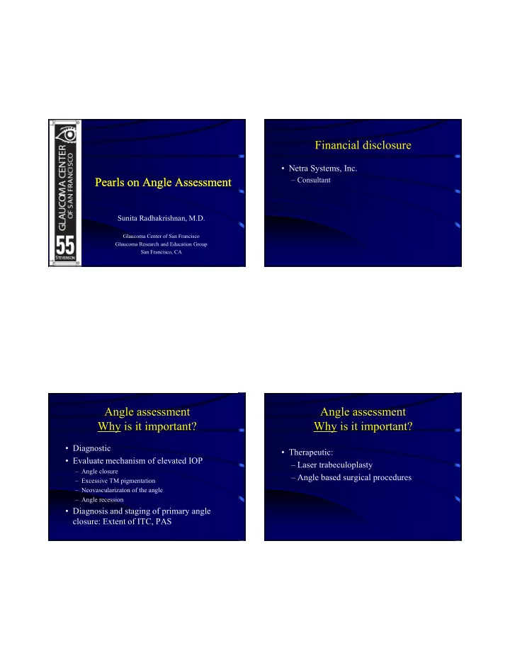
Pearls on Angle Assessment Pearls on Angle Assessment
Sunita Radhakrishnan, M.D.
Glaucoma Center of San Francisco Glaucoma Research and Education Group San Francisco, CA
Financial disclosure
- Netra Systems, Inc.
– Consultant
Angle assessment Why is it important?
- Diagnostic
- Evaluate mechanism of elevated IOP
– Angle closure – Excessive TM pigmentation – Neovascularizaton of the angle – Angle recession
- Diagnosis and staging of primary angle
closure: Extent of ITC, PAS
- Therapeutic: