
SLIDE 1
10 mm Cytoarchitecture and function layer 4: input layer 5: output - - PowerPoint PPT Presentation
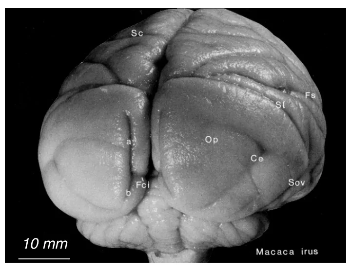
10 mm Cytoarchitecture and function layer 4: input layer 5: output Motor cortex: expanded layer 5, Primary visual cortex: expanded reduced layer 4 layer 4 with three sublayers Korbinian Brodmann ( 1868 - 1918 )
Primary visual cortex: expanded layer 4 with three sublayers Motor cortex: expanded layer 5, reduced layer 4 layer 4: input layer 5: output
Allman & Kaas, 1981 Zeki, 1978
Van Essen et al., 1992
V an Essen et al., 1992 V1 V3d V2d V3v V2v TH TF CITv AITv PITv PITd MT
CITd AITd STPa
FST
S T P p M S T l MSTd
V4d V4t V3A 7a DP LIP PIPVIP PO
MDP
M I P
Somato- sensory Orbito- frontal Lateral prefrontal Auditory Olfactory Motor 46 36 FEF Dorsal prefrontal Medial prefrontal Cingulate V4v ER ER HC LGN Retina SC M L I Pulvinar VOT 1 cm
PIT V1 V2 V3 PIP
V3a
MDP MIP PO MT V4 VIP LIP MST FST 7a STPp CIT STPa AIT O V T D P
W allisch & Movshon, 2008 after Felleman & V an Essen, 1991
Inflating and flattening the human cortex (Tootell and Dale)
fovea periphery horizontal meridian lower vertical meridian upper vertical meridian
fovea lower vertical meridian upper vertical meridian
Engel, Glover, & Wandell, Cereb Cortex (1997)
Brian Wandell
Brian Wandell
landmarks used to constrain the deformation. These include areas V1, V2, MT+, the central, Sylvian, and rhinal sulci, plus landmarks on the margins of cortex along the medial wall. Grid lines were carried passively with the deformation. (B) Landmarks and grid lines projected to the macaque spherical map. (C) Landmarks and grid lines deformed to the human spherical map. Neither of the spherical maps is at the same scale as the flat maps. (D) Deformed landmarks and grid lines projected to the human flat map. (E) Visual areas on the macaque flat map, based on the Lewis and Van Essen partitioning scheme in Fig. 4, plus iso-latitude and iso-longitude lines. (F) Visual areas on the macaque spherical map, plus iso-latitude and iso-longitude lines. (G) Deformed macaque visual areas on the human spherical map, along with deformed iso-latitude and iso-longitude lines. (H) Deformed macaque visual areas on the human flat map. To download these data, connect to http://stp.wustl.edu/sums/ sums.cgi?specfile=2001-03-06-VH.R.ATLAS–DeformedMa
V2 V3 V3A/B V7 IPS1 IPS2 V4 V5 (MT,MST) LO2 LO1
V1 V2 V3 V3A/B V7 IPS1 IPS2 V4 LO1 LO2 V5
Jonas Larsson and David Heeger
V an Essen et al., 1992 V1 V3d V2d V3v V2v TH TF CITv AITv PITv PITd MT
CITd AITd STPa
FST
S T P p M S T l MSTd
V4d V4t V3A 7a DP LIP PIPVIP PO
MDP
M I P
Somato- sensory Orbito- frontal Lateral prefrontal Auditory Olfactory Motor 46 36 FEF Dorsal prefrontal Medial prefrontal Cingulate V4v ER ER HC LGN Retina SC M L I Pulvinar VOT 1 cm
V an Essen, Anderson & Felleman, 1992
V1 V3d V2d V3v V2v TH TF CITv AITv PITv PITd MT
CITd AITd STPa
FST
S T P p M S T l MSTd
V4d V4t V3A 7a DP LIP PIPVIP PO
MDP
M I P
Somato- sensory Orbito- frontal Lateral prefrontal Auditory Olfactory Motor 46 36 FEF Dorsal prefrontal Medial prefrontal C i n g u l a t e V4v ER ER HC LGN Retina SC M L I Pulvinar VOT 1 cm
2/3 5/6 4 g a f e d c b k c a b d e e F d r a w r
d e e F 1
V1 V2 V4 TEO TEpd LIP 8m MST TH/TF peri 7A V3A V3 8L MT FST STPc 1 2 3 4 5 6 7 8 9 10
Level
DP
V an Essen, Anderson & Felleman, 1992; Markov et al., 2013
V1 V3d V2d V3v V2v TH TF CITv AITv PITv PITd MT
CITd AITd STPa
FST
S T P p M S T l MSTd
V4d V4t V3A 7a DP LIP PIPVIP PO
MDP
M I P
Somato- sensory Orbito- frontal Lateral prefrontal Auditory Olfactory Motor 46 36 FEF Dorsal prefrontal Medial prefrontal C i n g u l a t e V4v ER ER HC LGN Retina SC M L I Pulvinar VOT 1 cm
0.0 0.1 0.2 0.3 0.4 0.5 0.6 0.7 4.0 3.0 2.0 1.0 3.5 2.5 1.5
Graph density Average Pathlength
Modha and Singh 2010 Young 1993 Felleman and Van Essen, 1991 Jouve et al, 1998 predicted Jouve et al, 1998 Honey et al, 2007 Markov et al, 2013 FVE 1991 predicted
V1 V3d V2d V3v V2v TH TF CITv AITv PITv PITd MT
CITd AITd STPa
FST
S T P p M S T l MSTd
V4d V4t V3A 7a DP LIP PIPVIP PO
MDP
M I P
Somato- sensory Orbito- frontal Lateral prefrontal Auditory Olfactory Motor 46 36 FEF Dorsal prefrontal Medial prefrontal C i n g u l a t e V4v ER ER HC LGN Retina SC M L I Pulvinar VOT 1 cm
V an Essen, Anderson & Felleman, 1992; Markov et al., 2013
V an Essen, Anderson & Felleman, 1992
V1 V3d V2d V3v V2v TH TF CITv AITv PITv PITd MT
CITd AITd STPa
FST
S T P p M S T l MSTd
V4d V4t V3A 7a DP LIP PIPVIP PO
MDP
M I P
Somato- sensory Orbito- frontal Lateral prefrontal Auditory Olfactory Motor 46 36 FEF Dorsal prefrontal Medial prefrontal C i n g u l a t e V4v ER ER HC LGN Retina SC M L I Pulvinar VOT 1 cm
Adelson & Bergen, 1990
V1 V3d V2d V3v V2v TH TF CITv AITv PITv PITd MT
CITd AITd STPa
FST
S T P p M S T l MSTd
V4d V4t V3A 7a DP LIP PIPVIP PO
MDP
M I P
Somato- sensory Orbito- frontal Lateral prefrontal Auditory Olfactory Motor 46 36 FEF Dorsal prefrontal Medial prefrontal C i n g u l a t e V4v ER ER HC LGN Retina SC M L I Pulvinar VOT 1 cm
V an Essen, Anderson & Felleman, 1992; Markov et al., 2013
Niell, 2011
PIT V1 V2 V3 PIP
V3a
MDP MIP PO MT V4 VIP LIP MST FST 7a STPp CIT STPa AIT O V T D P
W allisch & Movshon, 2008 after Felleman & V an Essen, 1991
Physiological evidence for parallel cortical pathways? (Felleman and Van Essen, 1987)
Ungerleider & Mishkin, 1982
Object discrimination Landmark discrimination
Sir David Ferrier Lesions that caused blindness
Ungerleider & Mishkin, 1982
Milner & Goodale, 1995; Karnath et al 2009
Polar plots illustrating perceptual orientation judgements (A) and orientation adaptation in reaching movements (B). The photo inlays illustrate the respective tasks. The different
normalized to the vertical. The polar plots therefore show difference values to the vertical, representing a difference to the target orientation of 0°. Black data plots indicate the data of our patient J.S. and the data of VFA patient D.F. reported by Milner and Goodale (1995). Gray polar plots indicate an exemplary control of our study (A.K.) and the control subject reported by Milner and Goodale (1995) (Con). Bar plots illustrate SDs of J.S.'s responses in either task and average SDs in our group of healthy controls (error bars denote 1 SD).
typically seen as smaller than the central circle in the annulus of smaller circles, even though both central circles are actually the same size. (B) The same display, except that the central circle in the annulus of larger circles has been made slightly larger. As a consequence, the two central circles now appear to be the same size. (C) A 3-D version of the Ebbinghaus illusion. Participants are instructed to pick up one of the two 3-D disks placed either on the display shown in Panel A or the display shown in Panel B. (D) Two trials with the display shown in Panel B, in which the participant picked up the small disk on one trial and the large disk on another. Even though the two central disks were perceived as being the same size, the grip aperture in flight reflected the real not the apparent size of the disks. Adapted with permission from Aglioti et al. (1995).
Goodale, 2010
fMRI activation to intact versus scrambled line drawings. Note that the lesion (marked in blue) on patient D.F.’s right cerebral hemisphere encompasses all of area LO. Area LO in D.F.’s left hemisphere is also completely damaged. Adapted with permission from Goodale and Milner (2004).
Goodale, 2010
with optic ataxia, and DF, a patient with visual form agnosia. Panel A shows that RV was able to indicate the size of the objects reasonably well (individual trials marked as
DF showed excellent grip scaling, opening her hand wider for the 50 mm-wide object than for the 25-mm wide object. D.F.’s manual estimates of the width of the two objects, however, were grossly inaccurate and showed enormous variability from trial to trial.
Goodale, 2010
Adapted from John Maunsell
Nowak & Bullier, 1997
Stimulus (20 ms) ISI (30 ms) Mask (80 ms) + Animal present? ~50 ms SOA
Head Close-body Medium-body Far-body Animals Natural distractors Artificial distractors
Fabre-Thorpe, Richard & Thorpe, 1998
Stimulus (20 ms) ISI (30 ms) Mask (80 ms) + Animal present? ~50 ms SOA
Head Close-body Medium-body Far-body Animals Natural distractors Artificial distractors
Fabre-Thorpe, Richard & Thorpe, 1998
Stimulus (20 ms) ISI (30 ms) Mask (80 ms) + Animal present? ~50 ms SOA
Head Close-body Medium-body Far-body Animals Natural distractors Artificial distractors
Fabre-Thorpe, Richard & Thorpe, 1998
Cytochrome oxidase labelled stripes in a flattened section
V2.
Parallel visual pathways in macaque
Geniculate inputs to parallel visual pathways studied with laminar blockade
Maunsell, 1990