Vision III: Cortical mechanisms of First you tell them what youre - - PDF document

vision iii cortical mechanisms of first you tell them
SMART_READER_LITE
LIVE PREVIEW

Vision III: Cortical mechanisms of First you tell them what youre - - PDF document

Vision III: Cortical mechanisms of First you tell them what youre vision gonna tell them Please sit where you can examine a The cortical visual system is composed of multiple visual areas with different functions. partner. V1


slide-1
SLIDE 1

1

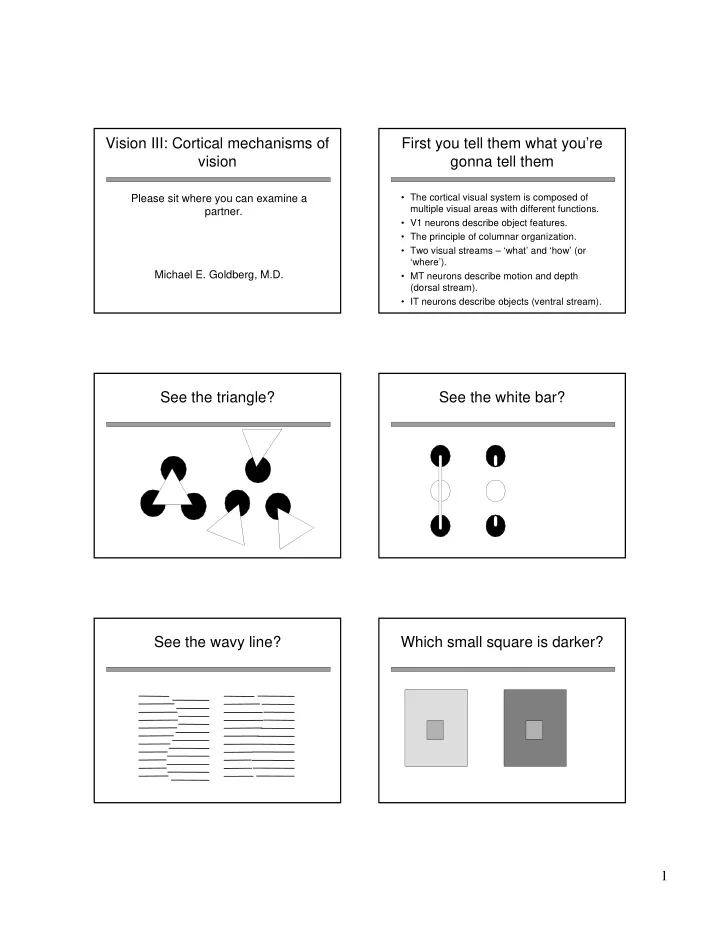
Vision III: Cortical mechanisms of vision

Please sit where you can examine a partner. Michael E. Goldberg, M.D.

First you tell them what you’re gonna tell them

  • The cortical visual system is composed of

multiple visual areas with different functions.

  • V1 neurons describe object features.
  • The principle of columnar organization.
  • Two visual streams – ‘what’ and ‘how’ (or

‘where’).

  • MT neurons describe motion and depth

(dorsal stream).

  • IT neurons describe objects (ventral stream).

See the triangle? See the white bar? See the wavy line? Which small square is darker?

slide-2
SLIDE 2

2

So

  • Your visual system does not measure

and report the exact physical nature of the visual world.

  • It collects some data, and makes

guesses.

  • Optical illusions take advantage of the

guessing strategies.

Roughly 40% of cerebral cortex is involved in vision

Off surround - inhibits

Remember

  • Receptive fields in the retina and the

lateral geniculate are circular, with center – surround organization.

On center - excites

The striate cortex – V1 – builds more sophisticated receptive fields from these basic building blocks. Cells describe specific

  • Contour orientations.
  • Binocular interaction.
  • Speed and direction of motion.
  • Color.

V1 simple cell is most responsive to an oriented line

Off-response On-response

Stimulus Angle (from max) Spikes/second

Orientation tuning in a V1 simple cell

slide-3
SLIDE 3

3

V1 complex cells are sensitive to

  • rientation of stimuli

But not particularly to stimulus position within the receptive field Complex cells can be constructed from an array of similarly oriented simple cells

geniculate nucleus I P P P M M 1 2 3 4A 4B 4Cα 4Cβ 5 Pyramidal Smooth stellate (local) 6 Blob Pyramidal Spiny stellate (local)

The cerebral cortex is organized in a columnar manner

To extrastriate Cortex – V2,V3 V4, MT To LGN, claustrum To SC,pulvinar pons

Within a column

  • Information is processed and transformed from

monocular, center-surround,non-directionally selective input to

  • Orientation-
  • Binocular disparity-
  • Direction-selective output
  • Processed information is distributed
  • Layers 2-3 to other cortical areas
  • Layer 5 to the superior colliculus
  • Layer 6 to the lateral geniculate nucleus
  • This general arrangement of columnar processing is

maintained throughout the cortex, not just visual cortex.

Cells with similar orientation preferences lie in the same column

slide-4
SLIDE 4

4 Geniculate cells representing the same area of the visual field but arising from different eyes project to adjacent areas

  • f V1

Orientation columns with the same monocular lateral geniculate input lie in the same ocular dominance column.

The actual topology of orientation and ocular dominance columns

Color sensitive cells lie at the center of the pinwheels, in cytochrome oxidase containing ‘blobs.’

Color sensitive cells are mostly unoriented

Depth perception starts with the detection of binocular disparity

A AR AL B BR BL C C C

Random dot stereograms generate structure from disparity

slide-5
SLIDE 5

5

Disparity selectivity in a V1 neuron Motion selectivity in a V1 neuron Two cortical visual streams subserve two different visual functions.

Where/how? What

Patients demonstrate this functional segregation

  • Patients with V1 lesions generally have total

visual field deficits in the affected field.

  • Patients with dorsal stream lesions have

deficits in sensory location (and attention), motion perception, color perception, and the performance of visually-guided movements.

  • Patients with ventral stream lesions have

visual agnosia, the inability to associate a visual stimulus with a name or function.

Functional separation begins in the retina and continues through the LGN

Retinal P cells: color, longer latency, fine detail LGN Parvocellular cells Retinal M cells: broadband, shorter latency courser detail LGN Magnocellular cells

And continues in V1

Blob Interblob

slide-6
SLIDE 6

6

V2 (Area 18) also is divisible by cytochrome oxidase staining

Stripes in Area 18 Blobs in Area 17

Functional separation continues in V2

After V2, different functions are performed by anatomically different areas: The dorsal stream provides vision for action –”where and how”

After V2, different functions are performed by anatomically different areas: The ventral stream provides vision for object identification

After V2, different functions are performed by anatomically different areas: But the areas are interconnected

MT – the analysis of motion

  • Neurons in MT are selective for speed

and direction of motion, and retinal disparity.

  • Neurons in MT report the perceptual

aspects of motion.

  • Electrical stimulation of MT affects the

perception of motion.

slide-7
SLIDE 7

7

Human MT Structure from motion MT Cells are tuned for direction Perceived motion in a plaid Striate neurons respond to the components of the plaid

Single component Plaid (2 components)

MT responds to the direction of the plaid, and not the components

Single component Plaid (2 components)

slide-8
SLIDE 8

8

MT has columns for direction of motion MT has disparity columns

Electrically stimulating an orientation column in MT induces the perception of motion described by that column

100% coherence

Electrically stimulating an orientation column in MT induces the perception of motion described by that column

50% coherence

Electrically stimulating an orientation column in MT induces the perception of motion described by that column

No coherence

Electrically stimulating an orientation column in MT induces the perception of motion described by that column

slide-9
SLIDE 9

9

The parietal lobe describes the world for action, location, and attention.

Where/how? What

There are multiple representations

  • f the visual field in the intraparietal

sulcus Within the dorsal stream there is further functional segregation –

  • MT is specialized for depth and motion.
  • LIP is specialized for attention in far

space.

  • MIP is specialized for providing visual.

information for reaching.

  • AIP is specialized for providing visual.

information for grasping.

  • VIP is specialized for providing visual.

information for mouth and head movements

An example of a dorsal stream function

  • When you reach for something, your

grip opens to accommodate the size of your target.

  • Patients with dorsal stream lesions can’t

do this.

  • They can, however, describe the size of

the object.

A patient with a dorsal stream lesion cannot orient her hand with respect to a slot

Neurons in AIP specialized for grip

Look at the

  • bject

Reach for the object

slide-10
SLIDE 10

10

The inferior temporal lobe describes the visual world for object recognition

Where/how? What?

Posting Matching

DF Control

A patient with a ventral stream lesion can move her hand to a slot, but can’t mimic the position.

Neurons in inferior temporal cortex are selective for complex patterns like faces

Copy the drawing Visuomotor function Intact – but patient can’t name the object Draw an anchor. Patient cannot conceptualize the anchor

Patients with inferior temporal lesions have visual agnosia Ventral stream patients

  • Cannot identify objects
  • But they can make appropriate visually-

guided movements.

  • The patient who could not set her grip

can still tell you which cylinder is thicker.

  • The patient who cannot tell you which

cylinder is thicker can set still her grip.

slide-11
SLIDE 11

11 Prosopagnosia “face blindness” is the most dramatic ventral stream deficit

  • Term first used by Bodamer, 1947
  • Inability to recognize familiar faces
  • Visual acuity is normal
  • Caused by lesion to right inferior temporal

lobe

  • May be congenital (“developmental

prosopagnosia”)

  • Patients compensate by using other

recognition cues: clothing, gait, voice, etc.

Finally, you tell them what you told them

  • The striate cortex (V1) uses unoriented, monocular

input from the lateral geniculate to assemble cells selective for orientation,motion, and retinal disparity. Complex cells generalize the orientation information found in simple cells.

  • Striate cortex is organized in columns with similar
  • rientation and ocular dominance.
  • Two visual streams emanate from V1: a dorsal

stream concerned with analyzing the visual world for location and action, and a ventral stream concerned with analyzing the nature of objects in the visual

  • world. Different areas subsume different spatial and
  • bject attribute functions.
  • Clinical deficits include specific deficits for color,

faces, motion, visual targeting of motion, and spatial