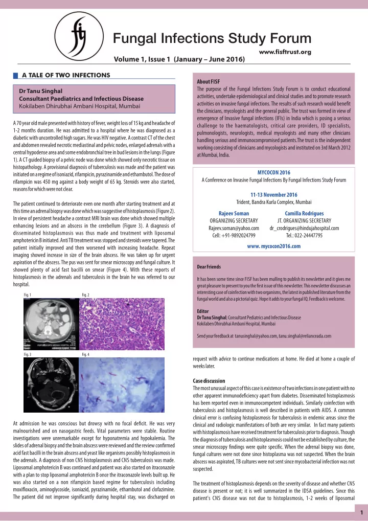

A 70 year old male presented with history of fever, weight loss of 15 kg and headache of 1-2 months duration. He was admitted to a hospital where he was diagnosed as a diabetic with uncontrolled high sugars. He was HIV negative. A contrast CT of the chest and abdomen revealed necrotic mediastinal and pelvic nodes, enlarged adrenals with a central hypodense area and some endobronchial tree in bud lesions in the lungs (Figure 1). A CT guided biopsy of a pelvic node was done which showed only necrotic tissue on
- histopathology. A provisional diagnosis of tuberculosis was made and the patient was
initiated on a regime of isoniazid, rifampicin, pyrazinamide and ethambutol. The dose of rifampicin was 450 mg against a body weight of 65 kg. Steroids were also started, reasons for which were not clear. The patient continued to deteriorate even one month after starting treatment and at this time an adrenal biopsy was done which was suggestive of histoplasmosis (Figure 2). In view of persistent headache a contrast MRI brain was done which showed multiple enhancing lesions and an abscess in the cerebellum (Figure 3). A diagnosis of disseminated histoplasmosis was thus made and treatment with liposomal amphotericin B initiated. Anti TB treatment was stopped and steroids were tapered. The patient initially improved and then worsened with increasing headache. Repeat imaging showed increase in size of the brain abscess. He was taken up for urgent aspiration of the abscess. The pus was sent for smear microscopy and fungal culture. It showed plenty of acid fast bacilli on smear (Figure 4). With these reports of histoplasmosis in the adrenals and tuberculosis in the brain he was referred to our hospital. At admission he was conscious but drowsy with no focal decit. He was very malnourished and on nasogastric feeds. Vital parameters were stable. Routine investigations were unremarkable except for hyponatremia and hypokalemia. The slides of adrenal biopsy and the brain abscess were reviewed and the review conrmed acid fast bacilli in the brain abscess and yeast like organisms possibly histoplasmosis in the adrenals. A diagnosis of non CNS histoplasmosis and CNS tuberculosis was made. Liposomal amphotericin B was continued and patient was also started on itraconazole with a plan to stop liposomal amphotericin B once the itraconazole levels built up. He was also started on a non rifampicin based regime for tuberculosis including moxioxacin, aminoglycoside, isoniazid, pyrazinamide, ethambutol and clofazimine. The patient did not improve signicantly during hospital stay, was discharged on request with advice to continue medications at home. He died at home a couple of weeks later. Case discussion The most unusual aspect of this case is existence of two infections in one patient with no
- ther apparent immunodeciency apart from diabetes. Disseminated histoplasmosis
has been reported even in immunocompetent individuals. Similarly coinfection with tuberculosis and histoplasmosis is well described in patients with AIDS. A common clinical error is confusing histoplasmosis for tuberculosis in endemic areas since the clinical and radiologic manifestations of both are very similar. In fact many patients with histoplasmosis have received treatment for tuberculosis prior to diagnosis. Though the diagnosis of tuberculosis and histoplasmosis could not be established by culture, the smear microscopy ndings were quite specic. When the adrenal biopsy was done, fungal cultures were not done since histoplasma was not suspected. When the brain abscess was aspirated, TB cultures were not sent since mycobacterial infection was not suspected. The treatment of histoplasmosis depends on the severity of disease and whether CNS disease is present or not; it is well summarized in the IDSA guidelines. Since this patient's CNS disease was not due to histoplasmosis, 1-2 weeks of liposomal
- Fig. 1
- Fig. 2
- Fig. 3
- Fig. 4
Dear Friends It has been some time since FISF has been mulling to publish its newsletter and it gives me great pleasure to present to you the rst issue of this newsletter. This newsletter discusses an interesting case of coinfection with two organisms, the latest in published literature from the fungal world and also a pictorial quiz. Hope it adds to your fungal IQ. Feedback is welcome. Editor Dr Tanu Singhal; Consultant Pediatrics and Infectious Disease Kokilaben Dhirubhai Ambani Hospital, Mumbai Send your feedback at tanusinghal@yahoo.com, tanu.singhal@relianceada.com
Dr Tanu Singhal Consultant Paediatrics and Infectious Disease Kokilaben Dhirubhai Ambani Hospital, Mumbai A TALE OF TWO INFECTIONS
Volume 1, Issue 1 (January – June 2016)
www.sftrust.org 1
Fungal Infections Study Forum
MYCOCON 2016 A Conference on Invasive Fungal Infections By Fungal Infections Study Forum 11-13 November 2016 Trident, Bandra Kurla Complex, Mumbai
- www. mycocon2016.com
About FISF The purpose of the Fungal Infections Study Forum is to conduct educational activities, undertake epidemiological and clinical studies and to promote research activities on invasive fungal infections. The results of such research would benet the clinicians, mycologists and the general public. The trust was formed in view of emergence of Invasive fungal infections (IFIs) in India which is posing a serious challenge to the haematologists, critical care providers, ID specialists, pulmonologists, neurologists, medical mycologists and many other clinicians handling serious and immunocompromised patients.The trust is the independent working consisting of clinicians and mycologists and instituted on 3rd March 2012 at Mumbai, India. Rajeev Soman ORGANIZING SECRETARY Rajeev.soman@yahoo.com Cell: +91-9892024799 Camilla Rodrigues
- JT. ORGANIZING SECRETARY
dr_crodrigues@hindujahospital.com Tel.: 022-24447795