7/24/2016 Plasma (Cell) Membrane Cell membrane separates living - - PDF document

7 24 2016
SMART_READER_LITE
LIVE PREVIEW

7/24/2016 Plasma (Cell) Membrane Cell membrane separates living - - PDF document

7/24/2016 Plasma (Cell) Membrane Cell membrane separates living cell from nonliving surroundings thin barrier = 8nm thick controls traffic in & out of the cell selectively permeable Chapter 7 allows some substances to cross


slide-1
SLIDE 1

7/24/2016 1

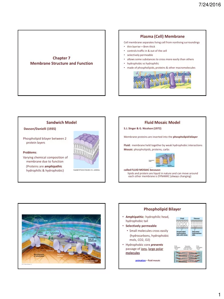
Chapter 7 Membrane Structure and Function Plasma (Cell) Membrane

Cell membrane separates living cell from nonliving surroundings

  • thin barrier = 8nm thick
  • controls traffic in & out of the cell
  • selectively permeable
  • allows some substances to cross more easily than others
  • hydrophobic vs hydrophilic
  • made of phospholipids, proteins & other macromolecules

Sandwich Model

Davson/Danielli (1935) Phospholipid bilayer between 2 protein layers Problems: Varying chemical composition of membrane due to function (Proteins are amphipathic hydrophilic & hydrophobic)

Fluid Mosaic Model

S.J. Singer & G. Nicolson (1972) Membrane proteins are inserted into the phospholipid bilayer Fluid: membrane held together by weak hydrophobic interactions Mosaic: phospholipids, proteins, carbs called FLUID MOSAIC because: lipids and protein are liquid in nature and can move around each other membrane is DYNAMIC (always changing)

Phospholipid Bilayer

  • Amphipathic: hydrophilic head,

hydrophobic tail

  • Selectively permeable
  • Small molecules cross easily

(hydrocarbons, hydrophobic mols, CO2, O2)

  • Hydrophobic core prevents

passage of ions, large polar molecules

animation – fluid mosaic

slide-2
SLIDE 2

7/24/2016 2

Polar vs Non-Polar Amino Acids

Nonpolar and hydrophobic AA

  • Interior of membrane
  • Anchors protein into membrane

polar area nonpolar areas Polar and hydrophilic AA

  • Outer surfaces
  • Extend into extracellular fluid

and into cytosol

Phospholipid Bilayer Structure

Freeze Fracture

Synthesis and Sidedness of Membranes

2 sides of membrane differ in specific lipid & protein composition Membrane is built by ER and Golgi apparatus

Membrane Fluidity

Low temps: phospholipids w/unsaturated tails (kinks prevent close packing) Cholesterol resists changes by: limit fluidity at high temps hinder close packing at low temps Adaptations: winter wheat  unsaturated phospholipids

  • fat composition affects

flexibility (like thick salad

  • il)

Membrane Proteins

Integral Proteins

Embedded in membrane Determined by freeze fracture Transmembrane with hydrophilic heads/tails and hydrophobic middles

Peripheral Proteins

Extracellular or cytoplasmic sides of membrane NOT embedded Held in place by the cytoskeleton or ECM (integrins) Provides stronger framework Act as identity markers (antigens)

Transmembrane Protein Structure

HYDROPHOBIC INTERIOR (alpha helix) HYDROPHILIC ENDS

slide-3
SLIDE 3

7/24/2016 3

Functions of Membrane Proteins

  • utside

Plasma membrane inside

  • 1. Transport

transport proteins:

  • involved in facilitated diffusion
  • go through the entire membrane
  • hydrophilic channel, hydrophobic outside
  • aquaporins: channel proteins increase water movement

2 types channel: gated pores move water and ions freely in and out of cell carrier: substrate binds to site on protein

  • 2. Cell-Cell Recognition

recognition proteins: (like tips of icebergs emerging from ocean surface)

  • Contain carbohydrate antennas (glycoproteins)
  • Used as chemical ID markers to differential cell types

***cell/cell recognition (immune response) ***embryo cells  tissues  organ systems *** A,B,O blood groups- carb antennae are different *** important in drug treatments

  • 3. Signal Transduction

receptor proteins: binds to specific molecules

  • specific shape binding site fits chemical messenger shape

(neurotransmitters, blood antigens, hormones)

  • initiates cell response
  • 4. Enzymatic Activity

enzymes: embedded in cell membrane to catalyze biochemical reactions in cell

  • 5. Intercellular Joining

Membrane proteins of adjacent cells hook together in various kinds of junctions

  • gap junctions: connects cytoplasm of cells

(cell- cell communicaiton)

  • tight junctions: fusion of adjacent cell membranes

(seal intercellular space, prevent free passage of substances)

slide-4
SLIDE 4

7/24/2016 4

  • 6. Attachment to Cytoskeleton and ECM

Microfilaments of cytoskeleton- non-covalently bound to membrane proteins

  • maintains cell shape
  • stabilizes location of certain membrane proteins
  • can coordinate extracellular and intracellular changes

Cell Transport- Passive Transport

  • 1. Diffusion
  • movement of molecules of a solute from areas of

high to low concentration (down a concentration gradient) until equilibrium is reached ex: hycrocarbons, CO2, Os, H2O

Cell Transport-Passive Transport

  • 2. Osmosis
  • movement of water from areas of high to low concentration

until equilibrium is reached

  • direction of movement depends on

CONCENTRATION OF WATER

  • n either side of the

concentration gradient ** differences in FREE WATER concentration is important **tonicity: ability of a surrounding solution to cause a cell to gain water

Effects of Osmotic Solutions

Osmoregulation: organisms lack rigid cell walls must control water and solute balance ex: contractile vacuole

Water Potential

Water potential (ψ)

  • free energy of water (potential energy)
  • predicts direction of water flow (solute concentration + physical

pressure)

  • H2O moves from high ψ  low ψ without any barrier to flow
  • Megapascal (Mpa) or Bars of pressure: unit of measurement

Ψ of pure water = 0 bars animation

Effects of solutes and pressure on water potential (ψ) and water movement

slide-5
SLIDE 5

7/24/2016 5

Pressure potential (ψP )

  • Physical pressure on a solution from pressure

inside or outside the cell (outward pressure of water in cell/ inward pressure of water

  • utside cell)
  • Can be positive or negative to atmospheric pressure

atmospheric pressure = 0 bars ψP ψP of open beaker of water = 0 bars animation Solute Potential (ψS)

  • smotic pressure
  • pressure that must be applied to a solution to prevent the inward

flow of water through a semi-permeable membrane

  • Solute always pulls water towards it
  • Inverse relationship with ψ (increase in solutes decreases free

water, therefore reduces water potential)

  • always expressed as 0 or a negative number

ψS pure water = 0 bars

Calculating Solute Potential (ψS)

ψS = -iCRT

i = ionization constant (how much particles ionize) always 1-2 C = molar concentration R = pressure constant (0.0831 liter bars/mole K) T = temperature in K (273 + 0C) bars = unit of measurement

The addition of solute to water lowers the solute potential (more negative) and therefore decreases the water potential animation

Water Potential Equation

Water potential equation: ψ = ψS + ψP Water potential (ψ) = free energy of water Solute potential (ψS) = solute concentration (osmotic potential) Pressure potential (ψP) = physical pressure on solution; turgor pressure (plants)

Pure water: ψP = 0 Mpa or bars Plant cells: ψP = 1 Mpa or bars

Which way will water move?

From an area of: higher ψ  lower ψ (more negative ψ) low solute concentration  high solute concentration high pressure  low pressure Example 1. Which chamber has a lower water potential? 2. Which chamber has a lower solute potential? 3. In which direction will osmosis occur? 4. If one chamber has a Ψ of -2000 kPa, and the other -1000 kPa, which is the chamber that has the higher Ψ?

slide-6
SLIDE 6

7/24/2016 6

How Does Water Move Up Plants? Sample Problem

1. Calculate the solute potential of a 0.1M NaCl solution at 25°C.

  • 2. If the concentration of NaCl inside the plant cell is 0.15M, which

way will the water diffuse if the cell is placed in the 0.1M NaCl solution?

Cell Transport- Passive Transport

  • 3. Facilitated Diffusion- carrier protein
  • type of passive transport
  • movement of a substance from areas of high to low

concentration (down gradient)

  • with the aid of a carrier protein (driven by diffusion)

ex: glucose or amino acids into RBC

Facilitated diffusion- gated ion channels

ion channel: transport protein that span

the thickness of the membrane with a polar pore through which ions can pass

  • allows ion to move thru membrane

without touching non polar lipid interior

  • form of passive transport: ions move

down concentration gradient

  • influenced by ions charge – the more

negative the charge, more likely to move

  • ut and vice versa
  • some gates always open
  • some gates stimulated by :

electrical charge stretching of membrane binding of specific molecules

Facilitated diffusion- channel protein

Aquaporin: channel protein that allows passage of H2O

Active Transport

  • movement of substances through a membrane against (up) a

concentration gradient (low  high concentration)

  • requires energy (from ATP)

ex: membrane pumps Na+/K+ pump proton (H+) pumps ex: bulk transport endo/exocytosis

slide-7
SLIDE 7

7/24/2016 7

Electrogenic Pumps: generate voltage across membranes

Na+/K+ Pump

  • Pump Na+ out, K+ into cell
  • Nerve transmission

animation

Proton Pump Push protons (H+) across membrane

  • mitochondria (ATP production)

animation

Bulk Transport

Endocytosis process where cells engulf substances too large to enter by passing through membrane Types

  • phagocytosis: cells engulf solid

particles too large to pass thru membrane

  • pinocytosis: cells engulf liquid

substances

  • receptor mediated:

ligands bind to specific receptors

  • n cell surface

Exocytosis process of removing large substances out

  • f cell

Ex: cells manufacture proteins vesicles fuse with cell membrane and dump contents

  • ut of cell
  • removal of cell debris,

bacteria/viruses, old organelle animation

Passive vs Active Transport

Passive

No ATP High  low concentration Down concentration gradient

Active

Requires ATP Low  high concentratin Against concentration gradient