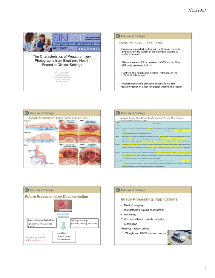

7/11/2017 Pressure Injury -- ‐ The Facts • Pressure is exerted on the skin, soft tissue, muscle and bone by the weight of an individual against a surface beneath. The Characteristics of Pressure Injury Photographs from Electronic Health • The incidence in ICUs between 1–56% and in Non- Record in Clinical Settings ICU units between 1--11%. Dan Li, Ph.D, RN • Costly to the health care system, total cost to the Assistant Professor U.S.=$11 billion/year. University of Pittsburgh School of Nursing dal144@pitt.edu • Require consistent objective assessments and documentation in order for proper treatment to occur. What Anatomical Locations are at Risk? *European Pressure Ulcer Advisory Panel and National Pressure Ulcer Advisory Panel (2016) Classification System Stage* Sign and symptoms ( 2016 ) Stage I Intact skin with a localized area of non -blanchable erythema, which may appear differently in darkly pigmented skin. Presence of blanchable erythema or changes in sensation, temperature, or firmness may precede visual changes. Color changes do not include purple or maroon discoloration; these may indicate deep tissue pressure injury. Stage II Partial-thickness loss of skin with exposed der mis. The wound bed is viable, pink or red, moist, and may also present as an intact or ruptured serum-filled blister. Adipose (fat) is not visible and deeper tissues are not visible. Granulation tissue, slough and eschar are not present. Full-thickness loss of skin, in w hich adipose (fat) is visible in the ulcer and granulation tissue and epibole (rolled wound edges) are often present. Stage III Slough and/or eschar may be visible. The depth of tissue damage varies by anatomical location; areas of significant adiposity can develop deep wounds. Undermining and tunneling may occur. Fascia, muscle, tendon, ligament, cartilage and/or bone are not exposed. If slough or eschar obscures the extent of tissue loss this is an Unstageable Pressure Injury. Stage IV Full-thickness skin and tissue loss with exposed or directly palpable fascia, muscle, tendon, ligament, cartilage or bone in the ulcer. Slough and/or eschar may be visible. Epibole (rolled edges), undermining and/or tunneling often occur. Depth varies by anatomical location. If slough or eschar obscures the extent of tissue loss this is an Unstageable Pressure Injury. Unstageable Full-thickness skin and tissue loss in which the extent of tissue damage within the ulcer cannot be confirmed because it is obscured by slough or eschar. If slough or eschar is removed, a Stage 3 or Stage 4 pressure injury will be revealed. Stable eschar (i.e. dry, adherent, intact without erythema or fluctuance) on the heel or ischemic limb should not be softened or remov ed. Future Pressure Injury Documentation Image Processing: Applications • Medical Imaging Tumor detection, wound assessment • Monitoring Traffic, surveillance, defects detection Image processing Technology Nursing knowledge (location, dressing, and odor) (granulation, color, size and • Automation stage ) Robotics, factory, driving Completed *Google and UBER autonomous car Pressure Injury A picture can be worth Documentation a thousand words! 1
7/11/2017 Applications: Digital Wound Assessment (DWA) Wound Assessment by Image Processing • Digital Wound Assessment Four Steps: (1) Preprocessing • Can be done locally or remotely (2) Segmentation (3) Image Analysis (4) Healing Projection • Can be 2D or 3D Why is Wound Photography Important? Image • 1. Allows for a formal record of pressure injury upon Processing admission Technology on • 2. Education for nursing and medical teams Pressure Injury Analysis • 3. Objective reproducible documentation • 4. Assessment of pressure Injury overtime How Should Photos be taken? How Should Photos be taken? --Wound Photography Protocol • Step 1: Prepare a digital camera with industry-standard resolution for high image quality • Step 2: Undress the wound and thoroughly cleanse the wound • Step 3: Position the camera perpendicularly to the wound • Step 4: Hold a small measurement grid flat along edge of the wound but not cover any part of wound • Step 5: Take the photographs under adequate light • Step 6: Upload the photos into the EHR 2
7/11/2017 Photography Characteristics Affecting Image Method Processing Wound Analysis • A 520-bed hospital in western Pennsylvania • Clinical background objects • 360 Pressure Injury Photographs from EHR Image processing wound assessment: Preprocessing • An experienced WOCN nurse and a nurse researcher • Relative position of the PI in the photographs reviewed all the PI photographs Image processing wound assessment: Segmentation • An image processing algorithm was used to calculate • Camera shooting angle camera shooting angle. Image processing wound assessment: Image Analysis Result: Statistics of Clinical Background Result: Quality of Pressure Injury Photographs Objects Clinical Number Percentage Variables Num ber Percentage Background Total collected 360 100% Objects photographs Bed linens 113 33.5% Gowns 155 46.0% Blurred 14 3.9% Other body parts 98 29.1% photographs Glove 56 16.6% Un-integrated PI 9 2.5% Ceiling and walls 47 13.9% Total qualified 337 93.6% Floor 69 20.5% photographs Others 86 26.4% Result: Statistics of pressure Injury relative position in the Result: Statistics of pressure Injury relative position in the images images 3
7/11/2017 Result: Theoretical Error of PI Surface Area Measurement Result: Statistics of Camera Shooting Angles in PI Photographs caused by Camera Shooting Angle 30% 1.2 26% 25% theoretical error on PI surface area measurement from PI image 23% 1 20% 19% 0.8 Frequency 15% 13% 0.6 11% 10% 0.4 5% 5% 0.2 2% 1% 0% 0% <10 ̊ 10 ̊ -20 ̊ 20 ̊ -30 ̊ 30 ̊ -40 ̊ 40 ̊ -50 ̊ 50 ̊ -60 ̊ 60 ̊ -70 ̊ 70 ̊ -80 ̊ 80 ̊ -90 ̊ 0 Camera shooting angle (Degree) 0 ̊ 10 ̊ 20 ̊ 30 ̊ 40 ̊ 50 ̊ 60 ̊ 70 ̊ 80 ̊ 90 ̊ Camera shooting angle (Degree) Discussions Conclusion • Photograph characteristics such as clinical background • The characteristics of pressure injury photos provide objects, camera angle, and the relative position of the PI preliminary evidence of how they affect image in the images do not affect wound assessment when processing and wound analysis. assessment from photographs by clinicians. • Certain standards and techniques must be followed • Image processing experts must consider clinical when photographing the PIs—or other chronic wounds background objects when developing image processing in order to further utilize the PI photographs. technologies for wound analysis. • Any method that is designed to retrieve wound dimension from wound photographs must incorporate a correction for suboptimal camera shooting angle. Thank you! 4
Recommend
More recommend