SLIDE 1
3/15/2017 1
17th Multidisciplinary Management of Cancers: A Case‐based Approach
Multidisciplinary Management of Cancers
Thoracic Oncology Tumor Board Session Chair Joel Neal, MD, PhD ‐ Stanford Assistant Professor of Medicine
17th Multidisciplinary Management of Cancers: A Case‐based Approach
Panel Members ‐ Deepti Behl, MD – Medical Oncology, Sutter Sacramento ‐ Colin Blakely, MD, PhD – Assistant Professor of Medicine, UCSF ‐ Lisa M. Brown, MD, MAS‐ Assistant Professor of Thoracic Surgery, UCDavis ‐ Megan Daly, MD – Assistant Professor of Radiation Oncology, UC Davis ‐ David Gandara, MD – Professor of Thoracic Medical Oncology, UC Davis ‐ Matthew Gubens, MD – Assistant Professor of Medicine, UCSF ‐ Billy Loo, MD, PhD, DABR – Associate Professor of Radiation Oncology, Stanford ‐ Joseph Shrager, MD – Professor of Cardiothoracic Surgery, Stanford ‐ Michael Mancuso, MD, PhD – Fellow in Oncology, Stanford
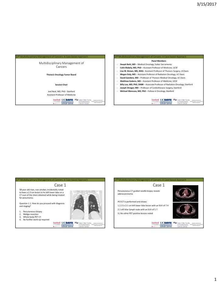
17th Multidisciplinary Management of Cancers: A Case‐based Approach
Case 1
58 year‐old man, non‐smoker, incidentally noted to have a 2.3 cm lesion in his left lower lobe on a CT scan of the chest obtained while being treated for pneumonia. Question 1.1. How do you proceed with diagnosis and staging? 1. Percutaneous biopsy 2. Wedge resection 3. Whole body PET‐CT 4. No further work‐up required
17th Multidisciplinary Management of Cancers: A Case‐based Approach
Percutaneous CT guided needle biopsy reveals adenocarcinoma PET/CT is performed and shows: 1.) 2.5 x 2.1 cm left lower lobe lesion with an SUV of 7.4 2.) Left hilar lymph node with an SUV of 5.7. 3.) No other PET positive lesions noted
Case 1
SLIDE 2 3/15/2017 2
17th Multidisciplinary Management of Cancers: A Case‐based Approach
Case 1
Question 1.2. Would you stage the mediastinum prior to lobectomy? If so, how? 1. Yes, Endobronchial Ultrasound and Biopsy 2. Yes, Mediastinoscopy 3. No (mediastinal lymphadenectomy with staged lobectomy)
17th Multidisciplinary Management of Cancers: A Case‐based Approach
The patient undergoes left VATS upper lobectomy with mediastinoscopy. PS = ECOG 1 after surgery. Pathology reveals invasive, moderately‐differentiated adenocarcinoma, measuring 2.5 cm, 1/12 lymph nodes positive for cancer (left hilar). Final pathology was pT1b pN1 (stage IIA, per TNM staging 7th edition; TNM 8th edition pT1c pN1, stage IIB). Margins are negative. While not currently standard of care in early stage disease, molecular testing is obtained and reveals EGFR L858R mutation.
Case 1
17th Multidisciplinary Management of Cancers: A Case‐based Approach
Updates to TNM staging of non‐small cell lung cancer (effective January 1, 2018)
T‐stage 7th edition 8th edition ≤ 1 cm T1a T1a > 1‐2 cm T1a T1b > 2‐3 cm T1b T1c > 3‐4 cm T2a T2a > 4‐5 cm T2a T2b > 5 cm‐7 cm T2b T3 Bronchi < 2 cm from carina T3 T2 Atelectasis/pneumonitis T3 T2 Invasion of diaphragm T3 T4 Invasion of mediastinal pleura T3 ‐ N component 7th edition 8th edition +/‐ LN involvement N0, N1, N2 N3 N0, N1, N2 N3 M component 7th edition 8th edition Metastasis within thoracic cavity M1a M1a Single extrathoracic metastasis M1b M1b Multiple extrathoracic metastasis M1b M1c
17th Multidisciplinary Management of Cancers: A Case‐based Approach
Descriptor in 7th edition T‐stage (8th edition) N categories Overall Stage 8th edition (7th edition)
N0 N1 N2 N3 T ≤ 1 cm T1a IA1 (IA) IIB (IIA) IIIA IIIB T1 > 1‐2 cm T1b IA2 (IA) IIB (IIA) IIIA IIIB T1 > 2‐3 cm T1c IA3 (IA) IIB (IIA) IIIA IIIB T2 > 3‐4 cm T2a IB IIB (IIA) IIIA IIIB T2 > 4‐5 cm T2b IIA (IB) IIB (IIA) IIIA IIIB T2 > 5‐7 cm T3 IIB (IIA) IIIA (IIB) IIIB (IIIA) IIIC (IIIB) T3 structures T3 IIB IIIA IIIB (IIIA) IIIC (IIIB) T3 > 7 cm T4 IIIA (IIB) IIIA IIIB (IIIA) IIIC (IIIB) T3 diaphragm T4 IIIA (IIB) IIIA IIIB (IIIA) IIIC (IIIB) T3 endobronchial location/atelectasis 4‐5 cm T2b IIA (IIB) IIB (IIIA) IIIA IIIB T4 T4 IIIA IIIA IIIB IIIC (IIIB) Upstaged from 7th to 8th edition Downstaged from 7th to 8th edition
SLIDE 3
3/15/2017 3
17th Multidisciplinary Management of Cancers: A Case‐based Approach
Question 1.3. Do you offer adjuvant therapy to this 58 year‐old man with no medical comorbidities and with a 2.5 cm adenocarcinoma of the lung with a positive hilar LN staged as TNM 7th edition pT1b pN1, stage IIA or TNM 8th edition pT1c pN1, stage IIB and negative margins after surgery? If so, what approach do you choose? 1. Adjuvant cytotoxic chemotherapy 2. Adjuvant radiation therapy 3. Adjuvant EGFR TKI therapy (such as erlotinib) 4. Adjuvant cytotoxic chemotherapy followed by adjuvant EGFR TKI therapy 5. No additional therapy
Case 1
17th Multidisciplinary Management of Cancers: A Case‐based Approach
Question 1.4. Which cytotoxic chemotherapy regimen would you recommend for this patient with 2.5 cm adenocarcinoma of the lung who is TNM 7th edition pT1b pN1, stage IIA or TNM 8th edition pT1c pN1, stage IIB? 1. Carboplatin‐Paclitaxel‐Bevacizumab 2. Carboplatin‐Pemetrexed 3. Cisplatin‐Pemetrexed‐Bevacizumab 4. Cisplatin‐Pemetrexed 5. Cisplatin‐Vinorelbine
Case 1
17th Multidisciplinary Management of Cancers: A Case‐based Approach
Question 1.5. Would your treatment be different if he had a positive margin after surgery? If so, what approach do you choose? He is a 58 year‐old man, with no medical comorbidities and with TNM 7th edition pT1b pN1, stage IIA or TNM 8th edition pT1c pN1, stage IIB disease (2.5 cm adenocarcinoma of the lung with a positive hilar LN). 1. Adjuvant chemotherapy only 2. Radiation therapy only 3. Sequential chemotherapy followed by radiation therapy 4. Sequential radiation therapy followed by chemotherapy 5. Combination chemoradiation therapy 6. Surgical re‐resection
Case 1
17th Multidisciplinary Management of Cancers: A Case‐based Approach
Question 1.6. If the lymph nodes were all negative for cancer, would you offer adjuvant cisplatin‐based chemotherapy for this patient if he were hypothetically TNM 7th edition Stage IA (pT1b pN0) or TNM 8th edition Stage IA3 (pT1c pN0) with a 2.5 cm adenocarcinoma? 1. Yes 2. No
Case 1
SLIDE 4 3/15/2017 4
17th Multidisciplinary Management of Cancers: A Case‐based Approach
Question 1.7. If the lymph nodes were negative for cancer and his tumor was 4.1 cm in size and staged as TNM 7th edition T2a N0 M0, stage IB or as TNM 8th edition T2b N0 M0, stage IIA, would you offer adjuvant cisplatin based chemotherapy if his surgical margin was negative? 1. Yes 2. No
Case 1
17th Multidisciplinary Management of Cancers: A Case‐based Approach
Question 1.8. Returning back to our 58 year‐old man with a left 2.5 cm adenocarcinoma with a positive left hilar lymph node, how frequently would you monitor this patient after completing therapy? What imaging modality would you choose in addition to H+P? 1. CT chest every 3‐6 months for 3 years and then every 6‐12 months through 5 years 2. #1 with alternating PET/CT 3. #1 with MRI brain annually 4. #1 with annual low dose chest CT after 5 years 5. #1 with regular chest CT every 1‐2 years for 10 years
Case 1
17th Multidisciplinary Management of Cancers: A Case‐based Approach
4 cycles of cisplatin/pemetrexed are completed. Follow‐up CT thorax at 6 months following surgery is clear.
Case 1
Follow‐up CT thorax at 12 months shows a new 13 mm lucent expansile lesion in the right rib and a new right pleural effusion. PET/CT also identifies an FDG avid right flank lesion MRI brain is negative
17th Multidisciplinary Management of Cancers: A Case‐based Approach
Case 1
Question 1.9. Do you obtain a biopsy of a new lesion? If so, do you also order repeat molecular testing (he has known EGFR L858R mutation in the original tumor)?
- 1. Obtain a tissue biopsy and order EGFR mutation testing
- 2. Obtain a tissue biopsy and do not order EGFR mutation testing
- 3. Proceed directly to treatment with targeted therapy given known history of EGFR L858R
mutated adenocarcinoma of the lung
SLIDE 5 3/15/2017 5
17th Multidisciplinary Management of Cancers: A Case‐based Approach
Case 1
Biopsy of the right flank region shows adenocarcinoma CK7+ TTF1+. PD‐L1 staining (22C3) is positive in 20% of cells The patient is started on erlotinib 150 mg daily with good response to therapy. Imaging 12 months later reveals progression of disease with new bone lesions at the sternum and right hip.
17th Multidisciplinary Management of Cancers: A Case‐based Approach
Case 1
Question 1.10. What is your next step in management?
- 1. Plasma or urine testing for EGFR T790M activating mutation
- 2. Obtain a tissue biopsy with repeat molecular testing for EGFR
- 3. #1, then if negative for T790M, #2 above
- 4. Continue erlotinib
- 5. Change therapy to cytotoxic chemotherapy
- 6. Change therapy to immunotherapy
17th Multidisciplinary Management of Cancers: A Case‐based Approach
Case 1
Molecular testing from the plasma identifies a new T790M mutation. Question 1.11. Which systemic therapy do you choose?
- 1. Osimertinib
- 2. Platinum + pemetrexed +/‐ bevacizumab
- 3. Carboplatin + paclitaxel +/‐ bevacizumab
- 4. Gefitinib
- 5. Afatinib
- 6. Pembrolizumab
- 7. Nivolumab
- 8. Atezolizumab
17th Multidisciplinary Management of Cancers: A Case‐based Approach
Case 1
Question 1.12. If the plasma and tissue based EGFR mutation assays were negative for T790M mutation, what would your next step be?
- 1. Osimertinib
- 2. Platinum + pemetrexed +/‐ bevacizumab
- 3. Carboplatin + paclitaxel +/‐ bevacizumab
- 4. Gefitinib
- 5. Afatinib
- 6. Pembrolizumab
- 7. Nivolumab
- 8. Atezolizumab
SLIDE 6 3/15/2017 6
17th Multidisciplinary Management of Cancers: A Case‐based Approach
Case 1: Take‐away points
- We are transitioning to the eighth edition of the TNM staging system in lung cancer
- Cisplatin based adjuvant therapy is still recommended for resected stage II and higher
NSCLC
- For EGFR positive NSCLC, testing should be done for the T790M resistance mutation,
which confers sensitivity to 3rd generation EGFR TKIs.
17th Multidisciplinary Management of Cancers: A Case‐based Approach
Case 2
ENT evaluation identifies vocal cord paralysis CT scan neck/chest with contrast showed 3 cm left apical mass with pathologically enlarged mediastinal and left hilar lymph nodes with likely involvement of the left recurrent laryngeal nerve 65 year‐old woman, who is a former smoker (40 pack year smoking history and quit 20 years ago) presents with one year hoarse voice and difficulty speaking.
17th Multidisciplinary Management of Cancers: A Case‐based Approach
Question 2.1. What is your next step in management?
- 1. Imaging with whole body PET/CT and MRI brain
- 2. Percutaneous biopsy of the primary mass
- 3. EBUS biopsy
- 4. Staged mediastinoscopy followed by lobectomy if negative
Case 2
17th Multidisciplinary Management of Cancers: A Case‐based Approach
PET/CT: 3.1 cm left upper lobe mass with SUV 14.9. Increased activity in left hilar and AP window lymph nodes with max SUV10.7. No evidence of metastatic disease.
Case 2
SLIDE 7 3/15/2017 7
17th Multidisciplinary Management of Cancers: A Case‐based Approach
CT‐guided biopsy of the lymph node mass reveals poorly differentiated adenocarcinoma (CK7 positive, TTF‐1 positive, CK20 negative). MRI brain is pending PET/CT scan is obtained
Case 2
17th Multidisciplinary Management of Cancers: A Case‐based Approach
Question 2.2. What therapy do you recommend for this patient with NSCLC TNM 7th edition clinical stage IIIA (T2a N2 M0) or Stage IIIA TNM 8th edition (T2a N2 M0)?
- 1. Induction chemoradiation surgery
- 2. Systemic therapy only
- 3. Chemotherapy surgery
- 4. Concurrent chemoradiation (without surgery)
- 5. Induction chemotherapy followed by radiotherapy
Case 2
17th Multidisciplinary Management of Cancers: A Case‐based Approach
A multidisciplinary plan is made, based on TNM 7th edition stage IIIA disease, to start therapy with cisplatin/etoposide (EP) 50/50 with concurrent IMRT radiation without plans for surgical resection.
Case 2
Prior to the start of therapy, MRI brain reveals multiple metastatic lesions and the plan to pursue chemoradiation therapy is deferred. Molecular testing is performed on the original biopsy specimen is performed: ‐ Negative for ALK and ROS1 rearrangements by FISH ‐ Negative for EGFR mutation
17th Multidisciplinary Management of Cancers: A Case‐based Approach
Question 2.3. What testing do you order to determine if the patient would benefit from first‐line immunotherapy with PD‐1 blockade?
- 1. Molecular testing for PD‐1 mutations
- 2. Molecular testing for PD‐L1 mutations
- 3. Immunohistochemical staining of PD‐L1 expression levels with antibody 22C3
- 4. Immunohistochemical staining of PD‐L1 expression levels with another antibody
- 5. No testing necessary
Case 2
SLIDE 8 3/15/2017 8
17th Multidisciplinary Management of Cancers: A Case‐based Approach
Approximately 70% of the cells in her biopsy specimen stain positive for PD‐L1 using the 22C3 antibody reagent. Question 2.4. What first line systemic therapy would you offer?
- 1. Platinum based chemotherapy
- 2. Pembrolizumab
- 3. Nivolumab
- 4. Atezolizumab
- 5. Chemotherapy + therapy targeting PD‐1/PD‐L1
Case 2
17th Multidisciplinary Management of Cancers: A Case‐based Approach
Question 2.7. If this patient were a non‐smoker and PD‐L1 IHC was still 70%, which therapy would you offer?
- 1. Platinum based chemotherapy
- 2. Pembrolizumab
- 3. Nivolumab
- 4. Atezolizumab
- 5. Chemotherapy + therapy targeting PD‐1/PD‐L1
Case 2
17th Multidisciplinary Management of Cancers: A Case‐based Approach
Patients with stage III NSCLC should be evaluated by a multidisciplinary team Chemotherapy, radiation, and surgery all can be part of the treatment of stage III NSCLC Pembrolizumab may be given front‐line for metastatic NSCLC if PD‐L1 expression positive (≥50%) and EGFR, ALK, ROS1 negative, regardless of smoking status
Case 2: Take‐away points
17th Multidisciplinary Management of Cancers: A Case‐based Approach
Case 3
55 year‐old woman, who has never smoked, presented with shortness of breath of two months duration and was found to have metastatic squamous cell carcinoma of the right lung. Molecular testing is negative for EGFR mutation and FISH is negative for ALK or ROS1 rearrangements. 10% of tumor cells express PD‐L1 (with antibody 22C3)
SLIDE 9 3/15/2017 9
17th Multidisciplinary Management of Cancers: A Case‐based Approach
Question 3.1. In this previously untreated advanced squamous non‐small cell lung cancer patient, which regimen would you give as first line therapy?
- 1. Platinum based chemotherapy (consider which doublet)
- 2. Therapy targeting PD‐1/PD‐L1
- 3. Chemotherapy + therapy targeting PD‐1/PD‐L1
Case 3
17th Multidisciplinary Management of Cancers: A Case‐based Approach
She receives 3 cycles of chemotherapy with carboplatin/paclitaxel with response Imaging after her 6th cycle of chemotherapy shows disease progression. PD‐L1 testing was 10% positive.
Case 3
Question 3.2. Which therapy do you choose in a patient with metastatic squamous cell carcinoma of the lung who has progressed after first‐line chemotherapy?
- 1. Docetaxel
- 2. Gemcitabine
- 3. Nab‐paclitaxel
- 4. Pembrolizumab
- 5. Nivolumab
- 6. Atezolizumab
17th Multidisciplinary Management of Cancers: A Case‐based Approach
She is started on nivolumab with excellent response as shown after 3 months.
Case 3
Questions 3.3. How often would you monitor this patient for response to therapy and for toxicity?
17th Multidisciplinary Management of Cancers: A Case‐based Approach
Case 3
After 6 months on nivolumab, laboratory studies show increase in serum aminotransferase levels Question 3.4. Given these findings, which course of action would you take?
- 1. Continue nivolumab
- 2. Continue nivolumab AND initiate
steroid therapy
- 3. Stop nivolumab and monitor
- 4. Stop nivolumab AND initiate steroid
therapy
ALT
SLIDE 10 3/15/2017 10
17th Multidisciplinary Management of Cancers: A Case‐based Approach
Case 3
Nivolumab is held and steroid therapy is initiated with improvement in her serum aminotransferase levels. Steroids are stopped and she is re‐started on nivolumab, but they worsen again.
ALT
Nivolumab held
Prednisone taper
Nivolumab given x1
Question 3.5. What is your next step in management?
- 1. Continue nivolumab AND
restart steroid therapy
- 2. Stop nivolumab and monitor
- 3. Stop nivolumab AND restart
steroid therapy
17th Multidisciplinary Management of Cancers: A Case‐based Approach
Case 3
Nivolumab continues to be held. Steroid dose is restarted and increased without immediate improvment Hepatology recommends mycophenolate mofetil if her AST/ALT continue to increase Fortunately, her AST and ALT normalize on higher doses of steroids
ALT
Nivolumab held
Prednisone taper Prednisone 60
Nivolumab given x 1
Prednisone 10
Nivolumab held
17th Multidisciplinary Management of Cancers: A Case‐based Approach
Case 3
While nivolumab is held due to her elevated AST and ALT, PET/CT scan shows development
- f a pleural based nodule as shown. The remainder of disease is still controlled.
Question 3.6. How do you manage this lesion?
- 1. Re‐initiate anti‐PD1 therapy
- 2. Radiation therapy to the lone site of
disease
- 3. Cytotoxic chemotherapy
17th Multidisciplinary Management of Cancers: A Case‐based Approach
Case 3: Take‐away points
Anti‐PD‐1/PD‐L1 therapies are active in metastatic squamous NSCLC in the first or second line setting Side‐affects of PD‐1/PD‐L1 inhibitors include autoimmune phenomena such as pneumonitis, hepatitis, colitis, adrenalitis, and thyroiditis Patients with autoimmune complications from PD‐1/PD‐L1 inhibitors can have improvement with steroids and considered for specialist referral
SLIDE 11 3/15/2017 11
17th Multidisciplinary Management of Cancers: A Case‐based Approach
Case 4
A 84 year‐old woman, never smoker, with hypertension, diabetes, and peripheral vascular disease presents with cough and is found to have a 3 cm solitary lung nodule on imaging. No additional evidence of disease is found on PET/CT. CT guided percutaneous biopsy shows adenocarcinoma of lung
17th Multidisciplinary Management of Cancers: A Case‐based Approach
Case 4
Question 4.1. What therapy do you recommend for an 84 year‐old patient with hypertension, diabetes, peripheral vascular disease and a 3 cm solitary ALK rearranged node negative NSCLC that is TNM 7th edition Stage IB (T2a N0 M0) or TNM 8th edition Stage IB (T2a N0 M0)?
- 1. Lobectomy or wedge resection
- 2. Lobectomy or wedge resection followed by adjuvant chemotherapy
- 3. Stereotactic radiation therapy
- 4. Stereotactic radiation therapy followed by adjuvant chemotherapy
- 5. Treatment with an ALK tyrosine kinase inhibitor
- 6. Treatment with Pembrolizumab
17th Multidisciplinary Management of Cancers: A Case‐based Approach
Case 4
She decides to pursue stereotactic radiation therapy. CT scans from before treatment and 3 months after therapy are shown.
Pretreatment scan 3 months after therapy
Question 4.2. For the three years after therapy, how is response monitored after radiation therapy to a solitary lesion? 1. CT ± contrast every 3‐6 months 2. CT ± contrast every 6 months with MRI brain every 12 months 3. PET CT every 3‐6 months 4. PET CT every 3‐6 months with MRI brain every 12 months
17th Multidisciplinary Management of Cancers: A Case‐based Approach
Case 5
51 year‐old woman presents with right upper quadrant pain and undergoes CT scan which shows CT chest 12.9 cm x 4.5 cm x 10 cm mass in the right costophrenic angle, multiple pleural‐based masses, and a large right pleural effusion. Question 5.1. What additional studies do you
- btain?
- 1. CT guided needle biopsy
- 2. VATS biopsy
- 3. No additional studies necessary,
mesothelioma is a clinical diagnosis
SLIDE 12 3/15/2017 12
17th Multidisciplinary Management of Cancers: A Case‐based Approach
Case 5
VATS biopsy reveals neoplastic cells positive for calretinin, CK mix, and CK5/6, negative for MOC31, p63, CD34 and S100. Findings consistent with malignant mesothelioma, deciduoid (epithelioid variant) type. Question 5.2. What treatment do you recommend?
- 1. Pleurectomy/decortication
- 2. Extrapleural pneumonectomy
- 3. Hemithoracic radiation therapy
- 4. Chemotherapy alone
- 5. Neoadjuvant chemotherapy followed by surgery
- 6. Chemotherapy and radiation
- 7. Trimodality therapy: neoadjuvant chemotherapy, surgery, adjuvant radiation
17th Multidisciplinary Management of Cancers: A Case‐based Approach
Case 5
Her case is presented at multidisciplinary tumor board and a plan is made for 3 cycles of neoadjuvant chemotherapy followed by repeat imaging to determine if the chest wall invasion regresses to make her a candidate for pleurectomy with decortication. Questions 5.3. What systemic therapy do you choose?
- 1. Cisplatin, pemetrexed
- 2. Cisplatin, pemetrexed, bevacizumab
- 3. Vinorelbine
17th Multidisciplinary Management of Cancers: A Case‐based Approach
Case 5
Questions 5.4. If she has a good response and proceeds with pleurectomy with decortication, what would be your next step in management?
- 1. Surveillance
- 2. Additional adjuvant chemotherapy
- 3. Adjuvant radiation therapy
- 4. Immunotherapy with anti‐PD‐1/PD‐L1
17th Multidisciplinary Management of Cancers: A Case‐based Approach
Case 5
She receives 3 cycles of cisplatin (75 mg/m2)/pemetrexed (500 mg/m2), with bevacizumab (15 mg/kg) added to cycles 1 and 2, with response to therapy as shown. She is ineligible for pleurectomy/decortication based on chest wall invasion.
Pretreatment scan Scan after 3 cycles of cis/pem/bev
SLIDE 13 3/15/2017 13
17th Multidisciplinary Management of Cancers: A Case‐based Approach
Case 5
Questions 5.5. If follow‐up imaging at 6 months shows disease recurrence, what second‐ line chemotherapy do you offer?
- 1. Gemcitabine
- 2. Anti PD‐1/PD‐L1 immunotherapy
- 3. Retreat with cisplatin + pemetrexed
- 4. Vinorelbine
She completes a total of 6 cycles of cisplatin/pemetrexed/bevacizumab with good response.
17th Multidisciplinary Management of Cancers: A Case‐based Approach
Case 5: Take‐away points
Malignant pleural mesothelioma has a median overall survival of 18‐36 months depending
- n performance status, stage, and histological sub‐type
First‐line chemotherapy for malignant pleural mesothelioma is platinum with pemetrexed, and optionally bevacizumab in eligible patients Patients who are surgical candidates should be referred to specialized centers with experience in mesothelioma to be considered for pleurectomy/decortication or extrapleural pneumonectomy as part of trimodality therapy
17th Multidisciplinary Management of Cancers: A Case‐based Approach
Thank you! Questions?