
SLIDE 1
1
UC SF
VASCULAR SURGERY • UC SAN FRANCISCO
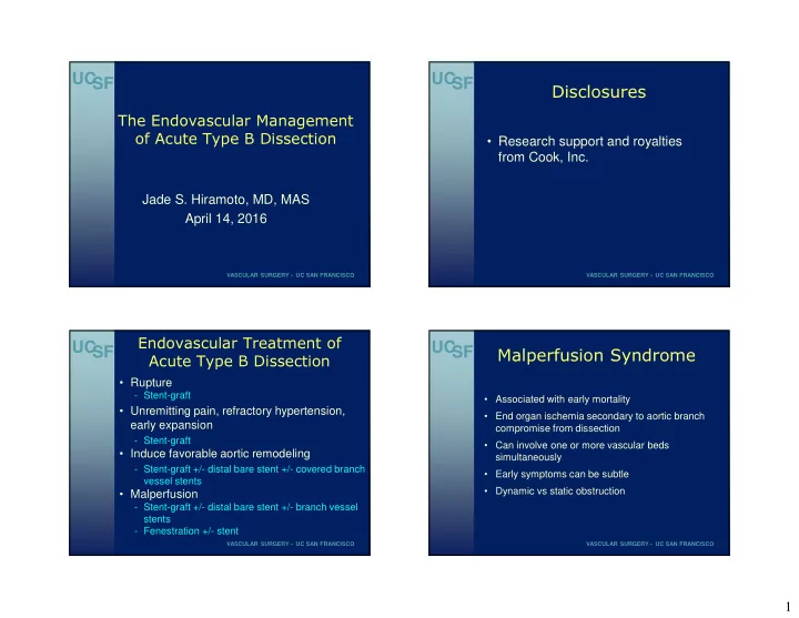
The Endovascular Management
- f Acute Type B Dissection
Jade S. Hiramoto, MD, MAS April 14, 2016
UC SF
VASCULAR SURGERY • UC SAN FRANCISCO
Disclosures
- Research support and royalties
from Cook, Inc.
UC SF
VASCULAR SURGERY • UC SAN FRANCISCO
Endovascular Treatment of Acute Type B Dissection
- Rupture
- Stent-graft
- Unremitting pain, refractory hypertension,
early expansion
- Stent-graft
- Induce favorable aortic remodeling
- Stent-graft +/- distal bare stent +/- covered branch
vessel stents
- Malperfusion
- Stent-graft +/- distal bare stent +/- branch vessel
stents
- Fenestration +/- stent
UC SF
VASCULAR SURGERY • UC SAN FRANCISCO
Malperfusion Syndrome
- Associated with early mortality
- End organ ischemia secondary to aortic branch
compromise from dissection
- Can involve one or more vascular beds
simultaneously
- Early symptoms can be subtle
- Dynamic vs static obstruction