The Blood-brain Barrier Dr. Eszter Farkas- Dr. Ferenc Domoki - - PowerPoint PPT Presentation

the blood brain barrier
SMART_READER_LITE
LIVE PREVIEW

The Blood-brain Barrier Dr. Eszter Farkas- Dr. Ferenc Domoki - - PowerPoint PPT Presentation

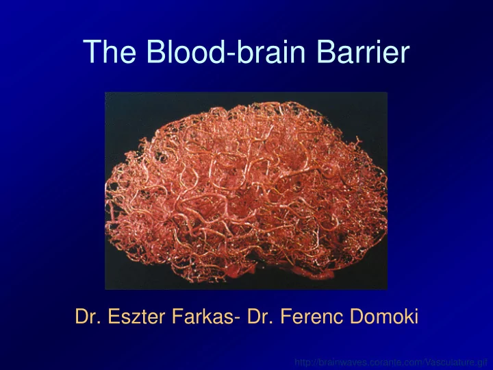
The Blood-brain Barrier Dr. Eszter Farkas- Dr. Ferenc Domoki http://brainwaves.corante.com/Vasculature.gif The discovery of the blood- brain barrier (BBB) Paul Ehrlich (1885): injection of colored dyes into the circulation all organs


slide-1
SLIDE 1

The Blood-brain Barrier

  • Dr. Eszter Farkas- Dr. Ferenc Domoki

http://brainwaves.corante.com/Vasculature.gif

slide-2
SLIDE 2

The discovery of the blood- brain barrier (BBB)

  • Paul Ehrlich (1885): injection of

colored dyes into the circulation → all organs were stained except for the central nervous system

  • Edwin Goldmann (1913): the dye was

injected into the central nervous system → the brain got stained, but no peripheral

  • rgans

⇒ proof for the existence of the BBB: the brain is an isolated

  • rgan
slide-3
SLIDE 3

The discovery of the blood-brain barrier: Paul Ehrlich and Edwin Goldman (1913)

Dye transport studies (Evans Blue-albumin, Na-fluorescein) are still in use and are important techniques to study the BBB integrity in vivo.

slide-4
SLIDE 4

The cerebral capillary network

  • Dense capillary network:

average intercapillary distance: 40μm

  • Large surface:

endothelial layer: 100cm2/g

  • Volume: the mass of

endothelial cells constitutes 0.1% of the brain tissue

http://www.teknat.uu.se/forskning/uu/bild.php?typ=forskningsprogram&id=225

slide-5
SLIDE 5

The cerebral capillary

Layers:

  • 1. Endothelium
  • 2. Basal membrane

(with pericytes)

  • 3. Astrocyte-endfeet

The tight junctions have been found to be responsible for the blocking of dye diffusion across the barrier

slide-6
SLIDE 6

The structure of the cerebral capillaries

slide-7
SLIDE 7

The ultrastructure of the cerebral capillaries

  • Endothelial cell (en: nucleus; ep: plasma; em: mitochondria)
  • Basal membrane (bm)
  • Pericyte (p)
  • Astrocytic endfeet (a)

Farkas & Luiten, Progr. Neurobiol. 2001

slide-8
SLIDE 8
  • P. Ballabh et al. / Neurobiology of Disease 16 (2004) 1–13

Molecular structure of the blood-brain barrier (BBB)

Tight junction: Primary seal is formed homodimers of claudins and occludin that are responsible for the low paracellular

  • permeability. Zonula
  • ccludens (ZO) proteins

anchor the seal to the cytoskeleton. Adherens junction: JAM and VE cadherins provide mechanical cell-to cell adhesion.

slide-9
SLIDE 9

Primary rat cerebromicrovascular endothelial cell and pericyte cultures

A B D C

50µm ZO-1 β-actin

  • ccludin

EC PC EC PC Rat EC PC EC PC Piglet

E

Domoki, F., et al.: Am J Physiol Reg Integr Comp Physiol 295:R1099-108, 2008.

slide-10
SLIDE 10

Transendothelial electrical resistance – a measure of the paracellular barrier in cell cultures

1DIV 2DIV 3DIV 4DIV 5DIV 50 100 150 200 250 1DIV 2DIV 3DIV 4DIV 5DIV 50 100 150 200 250 Rat CMVEC TEER (Ωcm2) Piglet CMVEC TEER (Ωcm2)

Domoki, F., et al.: Am J Physiol Reg Integr Comp Physiol 295:R1099-108, 2008.

slide-11
SLIDE 11

„In vitro BBB” modelling

Nakagawa S et al. Neurochem Res 54:253-263 (2009)

slide-12
SLIDE 12

blood brain endothelial cell

Transport throught the BBB: blood brain (influx)

Diffusion Simple Gases H2O Ethanol Facilitated Glucose Aminoacids Nucleosides Receptor-mediated endocytosis Ferro- transferin

×

  • - - -

+ + + + +

  • -

+ + + + + + + + + +

  • -

+ + + + +

  • Plasma proteins

Absorption-mediated endocytosis

slide-13
SLIDE 13

blood brain endothelial cell

Transport throught the BBB: brain blood (efflux)

S S S

P-glycoprotein (MDR1, ABCB1)

  • Pl. cancer

drugs G A N

glu gln gln glu gln

Na+ glutamine glutamate glu gln

glu gln gln glu

Glutamine secretion

slide-14
SLIDE 14

Endothelial enzyme and transport barrier: another feature of the blood-brain barrier

  • Many substances cannot pass the endothelial

cells because they are either (1) degraded by enzymes located in the luminal membrane or (2) pumped back to the blood by multispecific active transporters.

  • This barrier impedes greatly the delivery of

drugs into the central nervous system

  • The most important of such pumps are the P-

glycoprotein (multidrug resistant protein MDR1), the MDR-related protein (MRP2) and the organic anion transporter protein 2 (OATP2), they belong to a transporter superfamily: the ABC family!

slide-15
SLIDE 15

The ATP binding casette (ABC) transporter family

NBD: nucleotide binding domain TMD: transmembrane domain ABCB1=PGP

slide-16
SLIDE 16

ABCB1 (P glycoprotein) activity measurement - in vitro BBB

Note that polarity of ECs is maintained in the presence of pericytes and glial cells!

Nakagawa S et al. Neurochem Res 54:253-263 (2009)

slide-17
SLIDE 17

The enzymatic barrier

Enzyme Function Alkaline phosphatase De-phosphorylation (purine and pirimidine metabolism) Monoamino oxidase (MAO) Cathecolamine inactivation Aminopeptidase A Angiotensin metabolism Endopeptidase Break-down of neuropeptides (e.g. bradikynin, dynorphin, neurotensin) γ-glutamil transpeptidase Leukotriene C4 → D4 conversion

Enzymatic barrier: break-down of neuroactive substances

slide-18
SLIDE 18

Summary

Blood CSF Brain parenchyma

Tight junctions

slide-19
SLIDE 19

Areas outside the BBB

Function: – Hormone production

– Sensory function

– Production of CSF Circumventricular organs: – Pineal gland (3) – Median eminence – Neurohypophysis (5) – Subfornical organ (1) – Subcomissural organ (2) – Area postrema (4) – Organum vasculosum

  • f lamina terminalis (6)

– Choroid plexus

slide-20
SLIDE 20

endothelial cell blood brain

The opening of the blood brain barrier

×

Paracellular: increased permeability

  • f tight junctions

Transcellular: pinocytic transport

  • Pl. inflammation, ischemia, trauma

endothelial cell

slide-21
SLIDE 21

The opening of the blood-brain barrier

Structural correlate Conditions Mediators Loosening of the tight junction Hyperosmolarity, acidic pH, encephalitis, multiple sclerosis, ischaemia TNF-α, IL-β, histamin, bradykinin, serotonin, arachidonic acid, e.t.c. Pinocytic activity Hypertension, microwave irradiation, trauma, seizures, tumors TNF-α, IL-β, histamin, bradykinin, serotonin, arachidonic acid, e.t.c. Increased membrane fluidity Solvents (ethanol, propanol, buthanol, DMSO) Formation of pores Some antidepressants (chlorpromazine, notriptylin) Altered activation of transporters Diabetes, Alzheimer’s disease, stroke, obesitas, multiple sclerosis GLUT-1, ICAM-1

slide-22
SLIDE 22

The blood brain barrier: applications for medicine

2 big problems:

  • 1. Intact BBB function
  • 2. Impaired BBB function
slide-23
SLIDE 23

Ad 1: Drugs and the BBB

The intact BBB hinders/blocks drug delivery to the brain. It poses a problem for the treatment of many central nervous system diseases. Potential solutions: – Increased lipid-solubility of the drug – Transient opening of the BBB (e.g. osmotic) – „Wrapping” drugs (liposomes) – Intranasal pathway

slide-24
SLIDE 24
slide-25
SLIDE 25

Increased lipid solubility

  • Dihydropyridines: Ca2+channel antagonists
  • For the treatment of hypertension
  • Nimodipine: increased lipid solubility: for the treatment of

stroke

slide-26
SLIDE 26
slide-27
SLIDE 27

Osmotic opening of the blood-brain barrier

  • Intra-carotid infusion of hyperosmotic solutions (for

example: mannitol)

  • Transient shrinkage of the endothelial cells → loosening

and opening of the tight junctions

  • Defining variables:

– Length of infusion – Osmolarity of the solution

  • Influencing physiological parameters:

– Concentration of blood gases – Cardiac output

  • Therapeutical target: pl. chemotherapy of brain tumors
slide-28
SLIDE 28
slide-29
SLIDE 29

Application of liposomes

  • Known for 30 years

(Bangham)

  • Small, artificial

phospholipid vesicles

  • For medicines

targeting the CNS

  • In stroke treatment:

e.g. SOD

slide-30
SLIDE 30

Intranasal treatment strategies

  • Sensory nerve

endings: – n. olfactorius – n. trigeminalis

  • Nonisnvasive
  • NGF, IGF, FGF
  • Way of passage:
  • Intraneuronal: axonal transport, hours to days → for

specified brain regions

  • Extraneuronal: perineuronal, minutes → brain parenchyma,

CSF

slide-31
SLIDE 31

Ad 2: Impaired blood-brain barrier function

  • Amyloid angiopathy – Alzheimer’s disease
  • Basal membrane thickening – aging,

neurodegenerative disease, hypertension

  • (Atherosclerosis) – hypertension
  • Neuroinflammation (ischemia, trauma,

infection)

slide-32
SLIDE 32

Amyloid angiopathy

  • Amyloid precursor

protein: wrong slicing → β-amyloid peptide

  • Deposition into

vessel walls

  • Double ring: space

between the t. intima & media

  • Alzheimer’s disease
slide-33
SLIDE 33

Basal membrane thickening

  • Aging, dementia, hypertension
  • Hindered transport
slide-34
SLIDE 34

Atherosclerosis

  • Large vessels: rigidity, decreased flow
  • Small vessels: hindered flow & transport
slide-35
SLIDE 35

Inflammation at the BBB

  • causes: infection,

trauma, necrosis (stroke)

  • Inflammatory

mediators activate the contractile machinery

  • f the endothelial cells

severing the paracellular junctions. (signalling details next slide)

  • Permeability increases

+ cellular transmigration is made possible.

slide-36
SLIDE 36

Cellular signalling of EC contraction

slide-37
SLIDE 37

Abbreviations for the previous slide

  • TRPC1 – canonical transient receptor potential 1 ion channel (store-operated Ca2+

channel)

  • IP3, IP3R – inositol triphosphate, and its receptor (Ca2+ channel)
  • Gq, G12/13 – heterotrimeric G-proteins
  • PLC – phospholipase C
  • ER – endoplasmic reticulum
  • Rho – small G protein
  • RhoGEF – Rho guanin nucleotide exchange factor (activator of Rho)
  • ROK – Rho kinase
  • MYPT1 – myosine phosphatase targeting subunit 1
  • PP1 – protein phosphatase 1
  • CaM – calmodulin
  • MLC, MLCK – myosin light chain, MLC kinase
slide-38
SLIDE 38

Leukocyte trafficking through the blood vessel wall

  • On the surface of leukocytes: selectins, integrins (CAM families)
  • On the endothelial surface: other (containing immunglobulin domains)

CAMs (ICAM-1, ICAM-2, ICAM-3, VCAM) ⇒ Interaction

slide-39
SLIDE 39

Consequence of inflammatory BBB

  • pening
  • Elimination of pathogens, necrotic tissue
  • But also immune-mediated damage to

„innocent bystanders”

  • Edema formation that can impair blood

flow and transcapillary transport

  • Toxic metabolites or xenobiotics can

access the brain tissue

slide-40
SLIDE 40

Bone marrow stem cell transmigration through the blood-brain barrier

  • Female patients with leukemia
  • Bone marrow transplantation from a male relative
  • Examination of brain tissue
  • Cell clusters containing Y-chromosome in the brain
  • Most of them are glial cells
  • A small percentage of cells are neurons (7/10 000)
slide-41
SLIDE 41

Bone marrow stem cell transmigration through the blood-brain barrier

Mezey et al., PNAS, 2003. Green: NeuN Blue: nucleus Red: Y-chrom.

slide-42
SLIDE 42

Bone marrow stem cells: therapeutical potential for stroke?

  • Bone marrow transplantation from male mice to females,

male bone marrow cells express green fluorescent protein (GFP)

  • Middle cerebral artery occlusion → stroke
  • Histological examination of the brain:

labeling/visualization of GFP and Y-chromosomes

  • Labeled cells detected in the cerebral endothelial cells of

small vessels

  • Labeled, neuron-like cells in the brain
slide-43
SLIDE 43

Bone marrow stem cells: therapeutical potential for stroke?

Hess et al., Stroke, 2002.