
SLIDE 1
STEP 5. Slides of a neuron, nerve, and spinal cord #1 Neuron slide - - PDF document
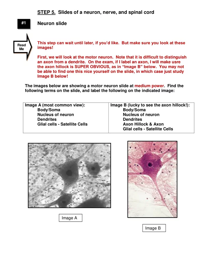
STEP 5. Slides of a neuron, nerve, and spinal cord #1 Neuron slide This step can wait until later, if youd like. But make sure you look at these Read images! Me First, we will look at the motor neuron. Note that it is difficult to