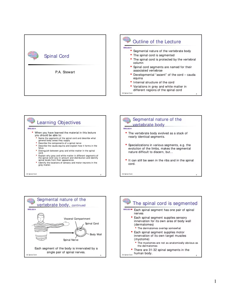

Outline of the Lecture • Segmental nature of the vertebrate body • The spinal cord is segmented Spinal Cord • The spinal cord is protected by the vertebral column • Spinal cord segments are named for their associated vertebrae P.A. Stewart • Developmental “ascent” of the cord – cauda equina • Internal structure of the cord • Variations in grey and white matter in different regions of the spinal cord 03 Spinal Cord 2 Segmental nature of the Learning Objectives vertebrate body • When you have learned the material in this lecture • The vertebrate body evolved as a stack of you should be able to: nearly identical segments. • Name the segments of the spinal cord and describe what general body areas they supply • Describe the components of a spinal nerve • Specializations in various segments, e.g. the • Describe the cauda equina and explain how it forms in the fetus. evolution of the limbs, makes the segmental • Distinguish between grey and white matter in the spinal nature difficult to discern, but... cord. • Explain why grey and white matter in different segments of the spinal cord vary in amount and distribution and identify • It can still be seen in the ribs and in the spinal spinal levels from their appearance. • Identify the locations of sensory and motor neurons in the cord. grey matter. 03 Spinal Cord 03 Spinal Cord 3 4 Segmental nature of the The spinal cord is segmented vertebrate body, continued • Each spinal segment has one pair of spinal nerves • Each spinal segment supplies sensory Visceral Compartment innervation for its own area of body wall Spinal Cord (dermatomes) • The dermatomes overlap somewhat • Each spinal segment supplies motor Body Wall innervation of its own target muscles (myotome) Spinal Nerve • The myotomes are not as anatomically obvious as the dermatomes Each segment of the body is innervated by a • There are 31-32 spinal segments in the single pair of spinal nerves. human body. 03 Spinal Cord 03 Spinal Cord 5 6 1
The spinal cord is protected by Vertebra – viewed from above the vertebral column Vertebral spine (the knobs you feel in your back) • The vertebral column is formed by a stack of Spinal canal bones called vertebrae. rib • Each vertebra has a body – a weight-bearing part, and a neural arch that houses the spinal Neural arch (houses cord spinal cord) • The neural arches of stacked the vertebrae forms the spinal (or vertebral) canal. Body (weight bearing) • The spinal nerves leave the spinal canal through foramena (openings) between the The grey areas are articular facets where the vertebrae. vertebra forms joints with the ribs and with vertebrae above and below 03 Spinal Cord 03 Spinal Cord 7 8 Spinal nerves are named for the Two Vertebrae viewed from the side vertebrae they are associated with Spinal cord Spinal nerves leave the spinal canal by passing • There are 7 cervical • There are 8(?) cervical Body between the vertebrae vertebrae (cervical spinal nerves. through an opening called • (a small spinal nerve, = neck) the intervertebral foramen • They are named C1- C1 emerges above (foramen = “window”) vertebrae C1) C7 starting at the • The remaining cervical top • Intervertebral discs form nerves, C2-C8 emerge cushions between vertebrae below vertebrae C1-C7 • They allow for flexibility in the spine • They can “rupture” and put pressure on a spinal nerve 03 Spinal Cord 03 Spinal Cord 9 10 Spinal nerves are named for the Spinal nerves are named for the vertebrae they are associated with vertebrae they are associated with • There are 12 thoracic • There are 12 thoracic • There are 5 sacral • There are 5 sacral vertebrae named T1- spinal nerves, named vertebrae named spinal nerves named T12 from the top down T1-T12, from the top S1-S5 S1-S5 down • Each spinal nerve is • There are 2 (or 3) • There are 2 (or 3) named for the vertebrae above which the nerve coccygeal vertebrae coccygeal spinal emerges • There are 5 Lumbar named Co1-Co 2(3) nerves named Co1- • There are 5 lumbar spinal nerves named Co2(3) vertebrae, named L1-L5 L1-L5 03 Spinal Cord 03 Spinal Cord 11 12 2
Distribution of the spinal nerves Dermatomes to the body • Nerves C1-C4 supply the neck • Nerves C5-T1 supply the upper limb • Nerves T1-L1 supply the trunk • Nerves L2-S2 supply the lower limb • The remaining nerves supply areas around the buttocks 03 Spinal Cord 03 Spinal Cord 13 14 Image from Netter, The Nervous System, CIBA 1953 The “ascent” of the cord The “ascent” of the cord • This has two During embryonic consequences: development the • The nerve roots elongate spinal nerves grow directly laterally and to stretch between their leave the spinal spinal segment and their canal in the foramen of exit. intervertebral foramen, but... • The bottom 1/3 of the The spinal canal spinal canal contains only grows faster and nerve roots and CSF. The longer than the bundle of nerve roots in spinal cord. the caudal canal are called the “cauda equina” From Moore & Persaud, 1998, The Developing Human, Page 459 From Moore & Persaud, 1998, The 03 Spinal Cord 03 Spinal Cord 15 16 Developing Human, Page 459 Cervical and lumbosacral Internal structure of the cord enlargements • Cervical segments (C5 –T1) and lumbosacral segments L2-S2 contain the sensory and motor neurons that supply the upper limbs. • Because the limbs have a large muscle mass and a high density of sensory endings, more motor and sensory neurons are located in these segments than in other areas of the cord. 03 Spinal Cord 03 Spinal Cord 17 18 3
Internal structure of the cord Internal structure of the cord • The white matter sleeve of the spinal cord is divided into three areas: the dorsal, lateral and ventral funiculi (funiculus = singular) • The dorsal funiculus is composed of ascending (sensory) axons carrying touch information from the body • The lateral funiculus is a mixture of ascending, sensory axons and descending, motor axons • The ventral funiculus consists almost exclusively of descending, motor axons 03 Spinal Cord 03 Spinal Cord 19 20 Internal structure of the cord Variations in White Matter • There is much more • The Grey matter core of the spinal cord is white matter at the butterfly-shaped in cross section. rostral end of the cord • It is divided into dorsal and ventral horns than at the caudal end • All of the descending motor axons destined for • The dorsal horn is composed of sensory neurons different segments re present at the rostral • The ventral horn is composed of motor neurons end. They terminate in various levels until, at the • In some regions of the cord (T1-L2), a lateral horn caudal end of the cord, there are none left is added. The lateral horn is composed of preganglionic sympathetic neurons. 03 Spinal Cord 03 Spinal Cord 21 22 Variations in White Matter Variations in White Matter Sacral level • Similarly, sensory High cervical level axons coming in from increasingly higher levels accumulate until, at the rostral end of the cord, all of the sensory axons are present. 03 Spinal Cord 03 Spinal Cord 23 24 4
Variations in Grey Matter Variations in Grey Matter • The dorsal and ventral horns extend the Midthoracic Level Cervical enlargement entire length of the spinal cord • In the cervical and lumbosacral enlargements – the areas that innervate the limbs - there are more motor and sensory neurons present.... • Therefore the grey matter is more massive at these levels 03 Spinal Cord 03 Spinal Cord 25 26 What level ? What level ? 03 Spinal Cord 03 Spinal Cord 27 28 What level ? What level ? 03 Spinal Cord 03 Spinal Cord 29 30 5
What level ? Summary of the Lecture • The vertebrate body and its spinal cord are segmented • Spinal cord segments are named for their associated vertebrae • During development the cord “ascends”. • The caudal spinal roots form the cauda equina • The spinal cord has a core of grey matter and a sleeve of white matter. • White matter increases in amount at higher levels of the cord • Grey matter increases in amount in the areas of the cord that innervate the limbs 03 Spinal Cord 03 Spinal Cord 31 32 6
Recommend
More recommend