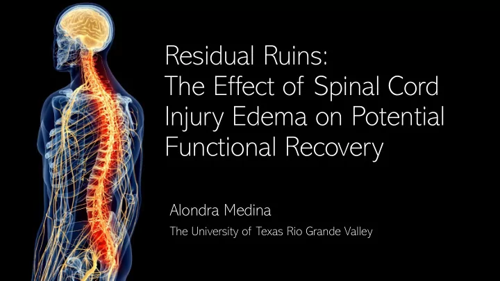

Residual Ruins: The Effect of Spinal Cord Injury Edema on Potential Functional Recovery Alondra Medina The University of Texas Rio Grande Valley
Spinal Cord Injury o Spinal cord injuries affect ~300,000 in the US o SCI cause varying degrees of motor and sensory impairment o Tetraplegia remains the most common SCI (>58%) o Currently over 9 months of rehabilitation are often required to achieve meaningful improvements in function in SCI 52 Muscle Strength of Chest (kg) 50 48 46 Functional 44 Recovery 42 40 38 Hicks, 2003 Duration of Therapy / Rehabilitation Training 36 Baseline 3 months 6 months 9 months
Spinal Cord Injury o Spinal cord injuries affect ~300,000 in the US o SCI cause varying degrees of motor and sensory impairment o Tetraplegia remains the most common SCI (>58%) o Currently over 9 months of rehabilitation are often required to achieve meaningful improvements in function in SCI 52 Muscle Strength of Chest (kg) 50 48 Can a neuroimaging biomarker assist in 46 Functional 44 determining what is causing limited Recovery 42 efficacy in rehabilitation? 40 38 Hicks, 2003 Duration of Therapy / Rehabilitation Training 36 Baseline 3 months 6 months 9 months
Spinal Cord Edema as a Biomarker o Following SCI, a series of scarring events occur around the zone of original damage o A cyst forms around the site of injury and becomes encapsulated by a glial scar
Spinal Cord Edema as a Biomarker o T2-weighted MRI imaging can be used to view the development of the edema o Over time, the edema compacts and stabilizes in size and shape Huber, 2017
Objective o The objective of our project was to determine if the properties of the residual edema in the spinal cord after SCI influenced functional recovery. o We hypothesized that subjects with a larger spinal edema would demonstrate limited recovery and reduced baseline function.
Methods Reh ehabil ilit itatio ion Neu euroimagin ing o Subjects underwent 10 sessions T2-weighted MRI of Edem ema Im Image Processing (2 hours each) the spinal cord o Intense upper limb rehabilitation o Upper limb function was assessed before and after therapy o Manual muscle strength testing and dexterity tests (nine hole peg test)
Methods o Image processing of edema and tissue bridges was performed in FSL o The spinal cord edema was first isolated as a separated region of interest (ROI) o Regions ventral and dorsal of the edema were also quantified (termed tissue bridges) o An example of the regions identified as tissue bridges are shown in yellow.
Methods Month ths Level of Le AIS Pati atient ID Gend nder Age Han andedn dness Post Etiology UE UEMS Inju njury Grad ade Inju njury K001 F 28 R 47 C5 B T 10 K002 M 68 R 135 C3 D T 38 K049 M 47 R 164 C4 D T 44 K092 F 67 R 417 C4 B T 23 K14 K146 M 57 R 30 C4 C T 25 K15 K153 F 32 R 79 C5 B T 22 K160 M 58 R 368 C6 D T 23 K207 M 56 R 60 C5 D T 33 o Patients were chronic SCI (over 18 months post injury) o All patients were incomplete tetraplegic (AIS Grade B,C, and D)
Edema Volume is Not Related to Baseline AIS grade 3500 Spinal Cord Edema Volume o We observed that subjects with varying 3000 baseline function had similar sized 2500 edemas regardless of baseline AIS 2000 (mm 3 ) grade. 1500 o This suggested that spinal cord edemas 1000 were not related to baseline function. 500 0 1 2 3 AIS B AIS C AIS AIS B AIS C AIS D
Edema Volume is not related to Recovery Potential 50% 45% 80% Change in Total Strength 40% 70% Change in Distal Strength 35% 60% r = . 297 30% 50% r = .542 25% 40% 20% 30% 15% 20% 10% 10% 5% 0% 0% 0 1000 2000 3000 4000 -10% 0 1000 2000 3000 4000 -20% Edema Volume Edema Volume o We found that total spinal cord edema volume did not correlate with recovery following two-weeks of rehabilitation.
Edema Volume is not related to Recovery Potential 50% 45% 80% Change in Total Strength 40% 70% Change in Distal Strength 35% 60% r = . 297 30% 50% r = .542 25% 40% 20% 30% 15% 20% 10% 10% 5% 0% 0% 0 1000 2000 3000 4000 -10% 0 1000 2000 3000 4000 -20% Edema Volume Edema Volume Edema volu lume was not t related to to baseline fu function o We found that total spinal cord edema volume did not correlate with recovery following two-weeks of rehabilitation. or r fun functional recovery
Tissue Bridge Volume is Dependent on AIS grade 1 0.9 0.8 Tissue Bridge Ratio 0.7 0.6 0.5 0.4 0.3 AIS B AIS C AIS D 0.2 0.1 o Sparing of the dorsal and ventral tissue bridges was 0 AIS B AIS C AIS D directly related to AIS grade. Ventral Dorsal o Our results validate clinical testing by demonstrating that individuals with AIS D showed the most sparing of both the ventral and dorsal tissue bridges.
Ventral Tissue Bridge Volume is Related to Recovery Potential 80% 50% Change in Distal Change in Total 40% 60% Strength Strength 30% 40% 20% r = .952 p = .003 10% 20% r = .930 p = .007 o Patients that demonstrated 0% 0% 0 1 2 3 a larger ventral tissue 0 1 2 3 Tissue Bridge Ratio: Tissue Bridge Ratio: bridge benefited more from Ventral Ventral rehabilitation. 100% Change in Distal 50% o Dorsal tissue bridge sparing Change in Total 80% 40% Strength had a trending relationship Strength 60% 30% with recovery. 40% 20% r = .770 p = .073 r = .794 p = .059 20% 10% 0% 0% 0 2 4 6 0 2 4 6 Tissue Bridge Ratio: Tissue Bridge Ratio: Dorsal Dorsal
Ventral Tissue Bridge Volume is Related to Recovery Potential 80% 50% Change in Distal Change in Total 40% 60% Strength Strength 30% 40% 20% r = .952 p = .003 10% 20% r = .930 p = .007 Patie tients ts with ith a lar larger 0% 0% 0 1 2 3 0 1 2 3 ve ventr tral tis tissue ue bri bridge Tissue Bridge Ratio: Tissue Bridge Ratio: Ventral Ventral demonstr trate ted mor ore 100% fu functional be benefit Change in Distal 50% Change in Total 80% 40% fol ollowing rehabilitati tion Strength Strength 60% 30% 40% 20% r = .770 p = .073 r = .794 p = .059 20% 10% 0% 0% 0 2 4 6 0 2 4 6 Tissue Bridge Ratio: Tissue Bridge Ratio: Dorsal Dorsal
Final Thoughts Can a neuroimaging biomarker assist in determining what is causing limited efficacy in rehabilitation? Ventral tissue bridges appear to provide the most feedback regarding rehabilitation efficacy
Future Research o Wider range of patients o Use MRI images from hospital and analyze more patients o Look at patients with AIS grade A o Find ways to enhance the survival of the ventral tissue bridge o Different stimulation techniques o Stem cells
Acknowledgements Cleveland veland Collab llaborator orators Ela Plow PhD, PT Kevin Kilgore, PhD Research team: Research partner: Dr. Kelsey Baker (Mentor), Juan Torres, Aaron Carrillo, Luis Alyssa Canales Trevino, Gisselle Montemayor, Rogelio Meza, Ileana Mendoza, Maria Martin, Carlos Arroyo, Claudia De Leon, Leslie Cardenas, and Nicole Alonzo Frederick Frost, MD Kyle O’Laughlin, MS Work Presented is in part of an active Clinical Trial: NCT01539109
Recommend
More recommend