Segmentation of blood vessels in retinal fundus images Healthy - - PowerPoint PPT Presentation

segmentation of blood vessels in retinal fundus images
SMART_READER_LITE
LIVE PREVIEW

Segmentation of blood vessels in retinal fundus images Healthy - - PowerPoint PPT Presentation

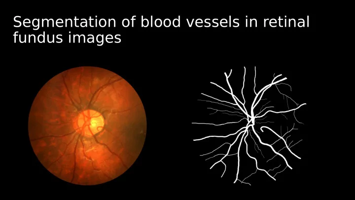
Segmentation of blood vessels in retinal fundus images Healthy Hypertension damage Ophtalmoscopy Retinal image Segmentation Automatic segmentation Simple bar-selective fjlter: B- COSFIRE Automatic confjguration f Each point


slide-1
SLIDE 1

Segmentation of blood vessels in retinal fundus images

slide-2
SLIDE 2
slide-3
SLIDE 3
slide-4
SLIDE 4

Healthy Hypertension damage

slide-5
SLIDE 5

Ophtalmoscopy

slide-6
SLIDE 6

Automatic segmentation Retinal image Segmentation

slide-7
SLIDE 7

Simple bar-selective fjlter: B- COSFIRE

Automatic confjguration Each point described by: Rotation invariance:

𝜍

f

slide-8
SLIDE 8

Filter application

  • Use a Gaussian for tolerance, std. Dev.:
  • Response for one point:
  • Multiply the shifted responses -> COSFIRE
slide-9
SLIDE 9

Pre-processing

Green channel Mask Original image

slide-10
SLIDE 10

Putting it all together

B-COSFIRE Threshold

slide-11
SLIDE 11

T uning parameters B-COSFIRE

  • σ:
  • ρ: The largest circle
  • σ0
  • α

𝜍

Symmetric: σ = 4.8, ρ = 20, σ0 = 3, α = 0.3 Assymetric: σ = 4.4, ρ = 36, σ0 = 1, α = 0.1

slide-12
SLIDE 12

Segmentation performance

t = [0,1] t TPR,FPR

slide-13
SLIDE 13

IOSTAR

EasyScan Optics B.V. The Netherlands

slide-14
SLIDE 14

Results

slide-15
SLIDE 15

Sensitivity of parameters

Paired T-test Small deviation Large performance difgerence Symmetric: σ0 = 3 σ = 4.8 α = 0.3 ed: Signifjcantly difgerent segmentation White: Similar segmentation

slide-16
SLIDE 16

Machine learning approaches

Training pase Working phase

slide-17
SLIDE 17

8 hours Single GPU 92 seconds High-end GPU ± 10 minutes Human 10 seconds 2 GHz CPU

Deep neural network B-COSFIRE

AUC: .9614 AUC: .9720

> >

>

Training/estimating Segmenting

slide-18
SLIDE 18

Thank you.