
SLIDE 1
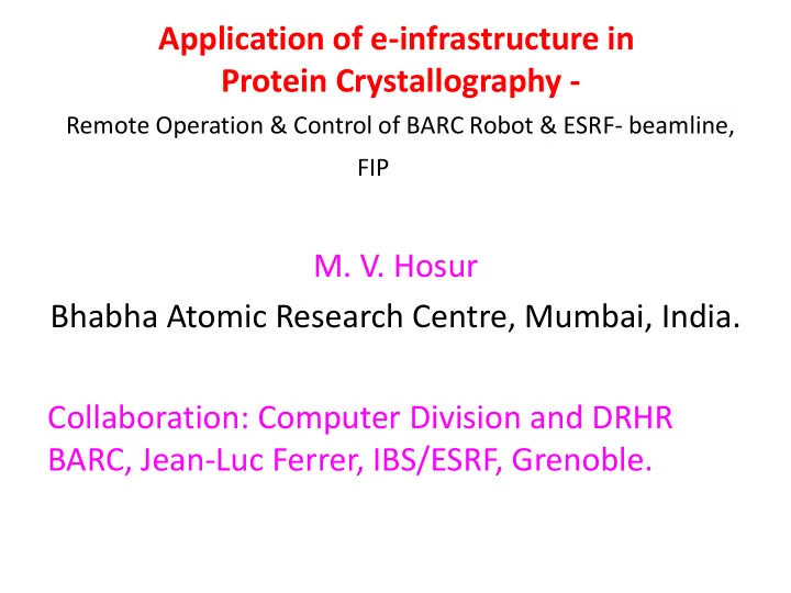
Application of e-infrastructure in Protein Crystallography - Remote Operation & Control of BARC Robot & ESRF- beamline,
FIP
- M. V. Hosur
Protein Crystallography - Remote Operation & Control of BARC - - PowerPoint PPT Presentation
Application of e-infrastructure in Protein Crystallography - Remote Operation & Control of BARC Robot & ESRF- beamline, FIP M. V. Hosur Bhabha Atomic Research Centre, Mumbai, India. Collaboration: Computer Division and DRHR BARC,
material.
acid-protein complexes.
[Fenn, Tanaka].
macromolecules in solution[Wüthrich].
Dramatic reduction in deaths due to AIDS is due to development of inhibitors of HIV enzymes through Structure-based drug-design approach. Has become a key component
has estimated a net annual gain of $2.4 trillion as a result of increased life expectancy alone through BR.
A typical oscillation pattern. Thousands
At different wave lengths for MAD. Repetitive nature of method.
A protein crystal(Many screened before selecting one)
X-rays
Crystal Diffracted X-rays
Commercial instruments do not provide for plate mounting
Can be transported to INDUS-II beamlines
ESRF – 6 GeV Grenoble, France, (I=1 million)
RAG – BARC, (I=1) LCLS Stanford (I=1 trillion)
Getting big crystals not always possible. Crystals as small as few microns can be used.
CATS robot Sample changer Sample storage cryo-dewar CCD X-ray detector X-ray beam Cryo-jet Crystal Single axis diffractometer
Softwares – usage for Nxclient – export desktop Visu – view exptal station Xnemo - Detector control, Input Data collection and storage parameters Gonio2 - Crystal mounting through CATS sample changer, crystal centering, mounting other tools Adxv - View diffraction image
Electron density map is contoured at 1σ. The four stranded β-sheet in HIV-1 protease/ritonavir drug complex is clearly visible. The inter-strand hydrogen bond lengths are marked.
Data collected by operating FIP beamline from HBNI, India. Resolution 1.6 Å. The data is of good quality , because, structure refined to low R (= 16.2%), and mutation is clearly seen. SA-OMIT map clearly shows electron density appropriate for the mutation M36I. The original amino acid Met (shown in purple carbons) doesn’t fit density. The changed amino acid Ile (shown in yellow carbons) fits the density perfectly.
N88S is resistant toward Nelfinavir. Data collected by operating FIP beamline from HBNI, India. Resolution 1.9 Å. structure refined to R (= 18.2%), Drug density is clearly seen. The drug is bound in two
Structure gives insight into drug- resistance mechanism.
Electron density for Nelfinavir
15 – 50 GeV. X-ray beam a billion times more intense than ESRF beams. Sample injected as 3 – 4 µl drops containing nano-crystals of proteins. Data collection at Room Temperature