
SLIDE 1
1
Definition, Diagnosis and Pathophysiology
Peggy Odegard, PharmD, BCPS, CDE
What face does “diabetes” bring to mind?
http://www.diabetes.org/diabetes statistics
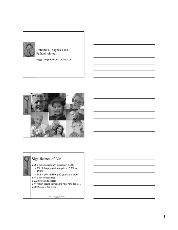
Significance of DM 20.8 million people with diabetes in the US 7% - - PDF document
Definition, Diagnosis and Pathophysiology Peggy Odegard, PharmD, BCPS, CDE What face does diabetes bring to mind? Significance of DM 20.8 million people with diabetes in the US 7% of the population (up from 5.9% in 1992)
http://www.diabetes.org/diabetes statistics
Diabetes Without Diabetes
$13,243 $2560 2000 4000 6000 8000 10000 12000 14000 Dollars
1000 2000 3000 4000 5000 6000 7000
Inpatient Nursing Home Physician's Office Outpatient Prescription Insulin and Supplies Home Health Hospital Outpatient Emergency Room
Dollars
Diabetes Without Diabetes Hogan P et al. Diabetes Care. 2003;26:917-932.
1) Stomach digests food 2) Liver stores glucose, then releases it later 3) Pancreas makes insulin
Glucose Metabolism Glucose Glucose Transporter Insulin Receptor Substrate Substrate-PO4 Protein synthesis Lipid synthesis Ion transport Cell growth Translocation
K+
ATP Channel
VDCC [ATP] [ADP] Insulin Glucose K+ Metabolism Depolarization ↑Free Ca++ Ca++
Glucose, nateglinide, repaglinide and sulfonylureas
Gly
1 5 1 5 10 15 20 S S 20 15 10
Gly Gln Ile Gln Cys Phe His His Leu
S S S S
Phe
25 30
ProLys Thr Ala
> 126 mg/dl < 126 mg/dl > 100 mg/dl < 100 mg/dl
70 mg/dl
Diabetes
IFG = Impaired Fasting Glucose; IGT = Impaired Glucose Tolerance
carbohydrate load
Diabetes
Cardiovascular disease
Disease (PVD)
Retinopathy Nephropathy Neuropathy Amputation
CDC National Diabetes Fact Sheet, November 2003.
Rohlfing CL et al. Diabetes Care. 2002;25:275; Bonora E et al. Diabetes Care. 2001;24:2023; Bastyr EJ et al. Diabetes Care. 2000;23:1236; Avignon et al. Diabetes Care. 1997;20:1822; De Veciana M et al. N Engl J Med. 1995;333:1237.
Glucose Red Blood Cell
CDC National Diabetes Fact Sheet, November 2003. National Eye Institute. Facts About Diabetic Retinopathy. Available at: www.nei.nih.gov/ health/ diabetic/ retinopathy.htm
CDC National Diabetes Fact Sheet, November 2003.
CDC National Diabetes Fact Sheet, November 2003.
CDC National Diabetes Fact Sheet, November 2003.
CDC National Diabetes Fact Sheet, November 2003.
CDC National Diabetes Fact Sheet, November 2003.
INTESTINE LIVER MUSCLE
Poor Food Choices and Obesity Decreased Insulin Production Insulin Resistance Increased Hepatic Glucose Production
Adapted from The Expert Committee on the Diagnosis and Classification of Diabetes Mellitus. Diabetes Care. 1997;20:1183-1197. 1-1
Genetic defects in β-cell function, exocrine pancreas diseases, endocrinopathies, drug- or chemical- induced, and other rare forms Other specific types Insulin resistance with β-cell dysfunction Gestational Insulin resistance with insulin deficiency Type 2 β-cell destruction with lack of insulin Type 1 Pathophysiology Classification
Type 1
♦ 10% of people with diabetes ♦ May start at any age (usually
<30 years of age)
♦ Rapid symptom onset ♦ Usually thin or lean ♦ Inability to produce insulin
(caused by destruction of insulin producing cells) Type 2
♦ 90% of people with diabetes ♦ Usually starts after age 30 ♦ Insidious onset ♦ 75% of patients are obese ♦ Caused by insulin resistance
insulin
Autoantibody negative Other forms If antibody positive, likely latent autoimmune diabetes in adults (LADA) with HLA similar to type1A Autoantibody negative Type 2 Rare in non-Hispanic white Autoantibody negative Type 1B Children:
♦90% non-Hispanic white ♦50% African American ♦50% Hispanic American
Autoantibody positive >90% Type 1A Comments Islet Antibodies Diabetes
– Islet cell autoantibodies (ICA) – Insulin autoantibodies (IAA) – Glutamic acid decarboxylase autoantibodies (GAD)
Glucose Metabolism Glucose Glucose Transporter Insulin Receptor Substrate Substrate-PO4 Protein synthesis Lipid synthesis Ion transport Cell growth Translocation
Adapted from Atkinson. Lancet. 2002;358:221-229.0
Age (years) Precipitating Event β-Cell Mass
Genetic predisposition Normal insulin release Glucose normal Overt diabetes No C-peptide present
Progressive loss
C-peptide present Antibody
American Diabetes Association. Clinical Practice Recommendations 2002. Diabetes Care. 2002;25(suppl 1):5.
50% 50% Monozygotic twin 10%-25% Father and Mother 6.5% 4.6% Father 5% 2% Mother 10% 6% Dizygotic twin 7.4% 3.2%, 6% lifetime Sibling 4.1% 1% Offspring 3% single Ab 0.3% multiple Ab 0.3% (15 – 25/ 100,000) General population (US) Islet Autoantibody % Childhood Diabetes (incidence/year) Proband with Diabetes
I m paired I nsulin Secretion I nsulin Resistance
I m paired I nsulin Secretion Glucose Toxicity I GT Hyperglycem ia of Type 2 Diabetes I nsulin Resistance I GT Glucose Toxicity
Kahn, et al. Diabetes. 1993;42:1663-1672.
Insulin Sensitivity Index (x 10-5 min-1/pM) Body Mass Index (kg/m
2)
15 20 25 30 35 40 45 50 55 30 25 20 15 10 5 Males Females
Fujimoto, et al. Obes Res. 1994;2:364-371.
Insulin Sensitivity Index (x 10-5 min-1/pM) Body Mass Index (kg/m2) r = .26 P =.35 Intra-abdominal Fat Area (cm2) r = .59 P <.05 10 20 22 24 26 28 30 32 4 2 6 8 10 20 22 24 26 28 30 32 4 2 6 8
Hyperglycemia
ADIPOSE ↑ Lipolysis ↑ FFA Mobilization
MUSCLE
↑ FFA Oxidation ↓ Glucose Utilization
LIVER
↑ FFA Oxidation ↑ Gluconeogenesis
Boden G. Proc Assoc Am Physicians. 1999;111:241
Kahn, et al. Diabetes. 1993;42:1663-1672
Insulin Secretion Insulin Sensitivity High Low Resistant Sensitive
95th 50th 5th Normal glucose tolerance IGT Type 2 diabetes
Adapted from Kahn, et al. Diabetes. 1993;42:1663-1672; Ward, et al. Diabetes. 1985;34:861-869; Kahn, et al. Am J Physiol. 1990; 258:E937-E943; Welch, et al. J Clin Endocrinol Metab. 1990;71:1508-1518; Ehrmann, et
Acute Insulin Response to Glucose (pM) Insulin Sensitivity Index (x 10 min
700 600 500 400 300 200 100 1 2 3 4 5 7 6 PCO women 50th 75th 25th 5th Older subjects Former GDM Relatives, type 2 diabetes Type 2 diabetes IGT
Robertson & Porte. J Clin Invest. 1973;52:870-876
Plasma Insulin (μU/mL) Time (min) Time (min) Normal Type2 Diabetes Glucos e 30 –30 40 20 60 80 100 30 –30 40 20 60 80 100 Glucos e
10 20 30 40 50 60 30 60 90 120 180 10 20 30 40 50 60 30 60 90 120 180 10 20 30 40 50 60 30 60 90 120 180
NGT Peak Insulin Response (% ) Time Post Oral Glucose Load (m in) I GT Peak Insulin Response (% ) Type 2 Diabetes Peak Insulin Response (% ) Time Post Oral Glucose Load (m in) Time Post Oral Glucose Load (m in) Frequency distribution by percentage of peak insulin after an oral glucose load Study sam ple subdivided by glucose tolerance status Bergstrom RW et al. J Clin Endocrinol Metab. 1990; 71: 1447
Polonsky, et al. N Engl J Med. 1988;318:1231-1239.
100 200 300 400
Glucose Insulin
0600 1000 1800 1400 0200 2200 0600 Time of Day 0600 1000 1800 1400 0200 2200 0600 Time of Day 20 40 60 80 100 120 B L S B L S
Normal Type 2 Diabetes
mg/dL μU/mL
6 7 8 9 1 2 3 4 5 6
A1C ( % ) Tim e ( yr)
Glycem ic Control
2 5 5 0 7 5 1 0 0 1 2 3 4 5 6
β-Cell Function
Tim e ( yr) β-Cell Function ( % ) Assessed by HOMA
UKPDS Group. Diabetes. 1995; 44: 1249 Diet/ Conventional Therapy Sulfonylurea
Every patient with type 2 diabetes eventually requires insulin.
Bagdade, et al. J Clin Invest. 1967; 46: 1549-1557.
Insulin (μU/mL) 250 200 150 100 50 15 30 45 60 90 120 150 180 Time (min) Normal (thin) DM (thin) Normal (obese) DM (obese)
Increase glycogenolysis and lipolysis Beta-2 agonists Increase insulin resistance by altering receptor-binding characteristics Atypical antipsychotics
1)
Inhibit insulin secretion due to hypokalemia
2)
Increased insulin resistance due to free fatty acids Thiazide diuretics Suppress conversion of proinsulin to insulin via calcium- dependent endopeptidases Protease inhibitors Inhibit insulin secretion Phenothiazines Increases insulin resistance due to increased free fatty acid mobilization Niacin Inhibit insulin secretion due to blockade of adenosine triphosphate (ATP)-sensitive potassium channels Fluoroquinolones Cause peripheral insulin resistance and gluconeogenesis Corticosteroids Inhibit insulin secretion due to inhibition of beta-cell cytosolic calcium Calcium-channel Blockers Inhibit insulin secretion (especially nonselective agents) Beta-blockers
Type 2 Diabetes I m paired I nsulin Secretion I nsulin Resistance I GT Genes Lifestyle Progressive Hyperglycem ia and High FFA
5.2 13 2 4 6 8 10 12 14 Million Patients Undiagnosed Diagnosed
CDC National Diabetes Fact Sheet, November 2003.
5 10 15 20 25 30 0-9 10-19 20-29 30-39 40-49 50-59 60-69 70-79 Age at Diagnosis of Diabetes Percent (%)
CDC National Diabetes Surveillance System, March 2005
14.9% 8.2% 11.4% 8.4% Percent of Patients in Ethnic Group 2 times as likely Asian Americans 2.2 times as likely 110,814 American Indians 1.5 times more likely* 2.0 million Hispanic/Latino Americans 1.6 times more likely 2.7 million Non-Hispanic Blacks 12.5 million Non-Hispanic Whites Relative Prevalence to Non-Hispanic Whites of Similar Age Number of Patients Race / Ethnicity * Mexican Americans are 2 times more likely; residents of Puerto Rico are 1.8 times more likely to develop diabetes than Non-Hispanic Whites.
CDC National Diabetes Fact Sheet, November 2003.
β-Cell Function ( % )
Postprandial Hyperglycem ia I GT Type 2 Diabetes Phase I Type 2 Diabetes Phase I I Type 2 Diabetes Phase I I I
25 10 0 75 50 –1 2 –1 0 –6 –2 2 6 10 14
Years From Diagnosis
Adapted from Lebovitz H. Diabetes Reviews. 1999; 7: 139
Alberti KGMM, Zim met PZ, for the WHO Consultation. Diabet Med. 1998; 15: 539
Expert Panel on Detection, Evaluation, and Treatment of High Blood Cholesterol in
♦ High-risk ethnic population
(e.g., African American, Latino, Native American, Asian American, Pacific Islander)
♦ First-degree relative with
diabetes
♦ Other clinical conditions
associated with insulin resistance (acanthosis nigricans)
♦ Polycystic ovary syndrome
(PCOS)
♦ Habitually physically inactive ♦ Hypertensive (140/90 mm Hg) ♦ HDL-C < 35 mg/dl and/or
TG > 250 mg/dl
♦ History of vascular disease ♦ History of IGT or IFG on
previous testing
♦ History of GDM or delivered a
baby weighing 9 lb
– BMI >85th percentile for age and sex – weight >85th percentile for height or >120% IBW
– FH of type 2 diabetes in 1st- or 2nd-degree relative – Race/ethnicity at high risk – Signs or conditions associated with insulin resistance (acanthosis nigricans, dyslipidemia, or PCOS)
– Age of initiation: age 10 years or at onset of puberty – Test: FPG preferred – Frequency: every 2 years
ADA: Clinical Practice Recommendations 2002. Diabetes Care. January 2002:25(suppl1) 5.
– <25 years of age – a normal body weight – no family history (i.e., first-degree relative) of diabetes – no history of abnormal glucose metabolism – no history of poor obstetric outcome – not members of an ethnic/racial group with a high prevalence of diabetes (e.g., Hispanic American, Native American, Asian American, African–American, Pacific Islander)
– Perform a diagnostic 100g glucose OGTT without prior plasma or serum glucose screening
1-h after a 50g oral glucose load
glucose threshold value 3-h Results >140 mg/dl are 80% sensitive for GDM; >130 mg/dl are 90% sensitive for GDM
100g OGTT 140 3-h 155 2-h 180 1-h 95 Fasting Glucose mg/dl Time