Objectives Multiple Myeloma Understand the eligibility and purpose - - PDF document

objectives multiple myeloma
SMART_READER_LITE
LIVE PREVIEW

Objectives Multiple Myeloma Understand the eligibility and purpose - - PDF document

Objectives Multiple Myeloma Understand the eligibility and purpose and MGUS of hematopoietic stem cell transplantation for myeloma. Craig Hofmeister, M.D. Understand who will benefit from intravenous bisphosphonates and the Assistant


slide-1
SLIDE 1

1

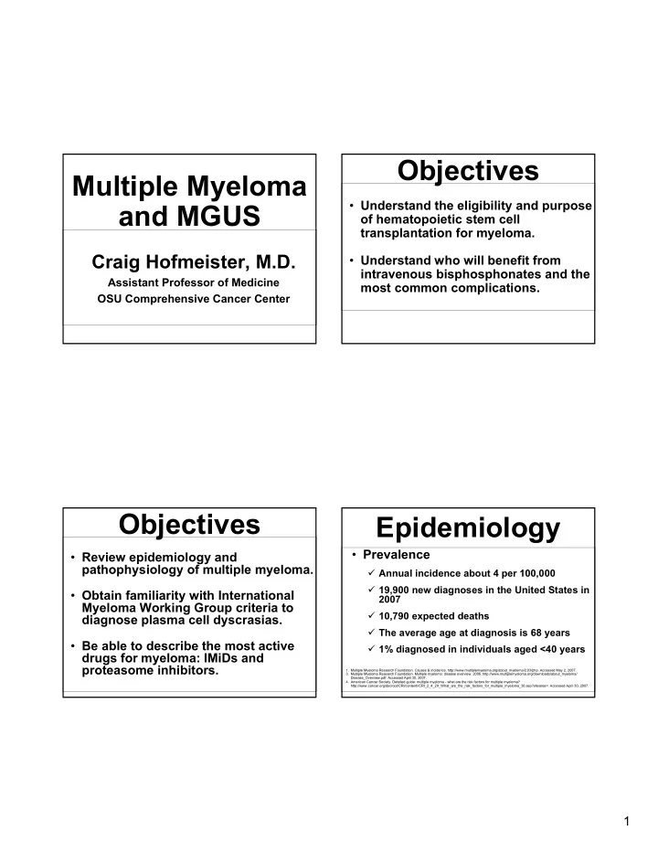
Multiple Myeloma and MGUS

Craig Hofmeister, M.D.

Assistant Professor of Medicine OSU Comprehensive Cancer Center

  • Review epidemiology and

pathophysiology of multiple myeloma.

  • Obtain familiarity with International

Myeloma Working Group criteria to diagnose plasma cell dyscrasias.

  • Be able to describe the most active

drugs for myeloma: IMiDs and proteasome inhibitors.

Objectives

  • Understand the eligibility and purpose
  • f hematopoietic stem cell

transplantation for myeloma.

  • Understand who will benefit from

intravenous bisphosphonates and the most common complications.

Objectives

  • Prevalence

Annual incidence about 4 per 100,000 19,900 new diagnoses in the United States in 2007 10,790 expected deaths The average age at diagnosis is 68 years 1% diagnosed in individuals aged <40 years

Epidemiology

  • 1. Multiple Myeloma Research Foundation. Causes & incidence. http://www.multiplemyeloma.org/about_myeloma/2.03/php. Accessed May 2, 2007.
  • 3. Multiple Myeloma Research Foundation. Multiple myeloma: disease overview. 2006. http://www.multiplemyeloma.org/downloads/about_myeloma/

Disease_Overview.pdf. Accessed April 30, 2007.

  • 4. American Cancer Society. Detailed guide: multiple myeloma - what are the risk factors for multiple myeloma?

http://www.cancer.org/docroot/CRI/content//CRI_2_4_2X_What_are_the_risk_factors_for_multiple_myeloma_30.asp?sitearea=. Accessed April 30, 2007.

slide-2
SLIDE 2

2

  • Population subgroups

Incidence of multiple myeloma (MM) is twice as common in African Americans Slightly more frequent in men than women

  • 10,960 men; 8940 women (estimated new

cases in 2007)

  • Remains incurable

Epidemiology

  • 1. Multiple Myeloma Research Foundation. Causes & incidence. http://www.multiplemyeloma.org/about_myeloma/2.03/php. Accessed May 2, 2007.
  • 3. Multiple Myeloma Research Foundation. Multiple myeloma: disease overview. 2006. http://www.multiplemyeloma.org/downloads/about_myeloma/

Disease_Overview.pdf. Accessed April 30, 2007.

  • 4. American Cancer Society. Detailed guide: multiple myeloma - what are the risk factors for multiple myeloma?

http://www.cancer.org/docroot/CRI/content//CRI_2_4_2X_What_are_the_risk_factors_for_multiple_myeloma_30.asp?sitearea=. Accessed April 30, 2007.

Epidemiology

Age-specific Incidence Rates for Myeloma, 2000–2003

*<25 cases for this age group. Source: SEER Cancer Statistics Review 1975-2003, National Cancer Institute, 2006.

50 40 30 20 10

Incidence (per 100,000)

85+ 80-84 75-79 65-69 70-74 60-64 55-59 50-54 40-44 45-49 35-39 30-34 0-24*

Age in Years

25-29

  • Common clinical features include

Bone pain, fractures, osteolytic lesions Anemia, hypercalcemia Renal insufficiency Bleeding tendency Peripheral neuropathy

Epidemiology

  • 1. American Cancer Society. Cancer facts & figures. 2002. 2003. 2004. 2005. 2006. 2007. http://www.cancer.org. Accessed April 30, 2007.
  • 2. International Myeloma Foundation. Concise review of the disease and treatment options. 2006.

http://www.myeloma.org/pdfs/ConciseReview2006.pdf. Accessed April 30, 2007.

10,900 14,600 2003 10,800 14,600 2002 2007 2006 2005 2004 10,790 11,310 11,300 11,070 Deaths 19,900 16,570 15,980 15,270 New Cases

Estimated Annual Cases in the United States

Clinical Presentation

  • Bone pain
  • Bone marrow

Easy bruising Infection Anemia

  • Headaches
  • Blurry vision
  • Renal insufficiency
  • Hypercalcemia

Somnolence Nausea and vomiting

  • 1. International Myeloma Foundation. Concise review of the disease and treatment options. 2006.

http://www.myeloma.org/pdfs/ConciseReview2006.pdf. Accessed April 30, 2007.

  • 2. Multiple Myeloma Research Foundation. Multiple myeloma: disease overview. 2006.

http://www.multiplemyeloma.org/downloads/about_myeloma/Disease_Overview.pdf. Accessed April 30, 2007.

slide-3
SLIDE 3

3

  • Hematologic

73% of patients will have anemia secondary to

  • Marrow infiltration with plasma cells
  • Decreased levels of erythropoietin
  • Decreased erythrocyte survival

Systems Affected

Kyle RA, et al. Mayo Clin Proc. 2003;78:21-33.

  • Skeletal

Bone pain is the most common symptom

  • Osteolytic lesions, generalized
  • steoporosis, or osteopenia

Associated oncologic emergencies

  • Spinal cord compression
  • Hypercalcemia (Not a true emergency)
  • Renal

M proteins may lead to renal failure

Systems Affected

Kyle RA, et al. Mayo Clin Proc. 2003;78:21-33.

Renal Manifestations

  • 55%: Myeloma kidney = Cast

nephropathy, i.e. tubular casts in the distal nephron

  • 20% AL amyloidosis

in glomeruli and blood vessels

  • 20% Monoclonal Ig deposition

disease, most commonly light-chain deposition disease

  • 5% Cryoglobulinemic glomerulonephritis &

proliferative glomerulonephritis (rare)

Congo red birefringence of a glomerulus when viewed through crossed Polaroid filters

In MM, there is overproduction of one subtype

  • f immunoglobulin, called the M protein

(monoclonal protein)

“M-spike”

Adapted from International Myeloma Foundation. Concise review of the disease and treatment options. 2006. http://www.myeloma.org/pdfs/ConciseReview2006.pdf. Accessed April 30, 2007.

slide-4
SLIDE 4

4

Serum Protein Electropheresis +

Control Patient

albumin alpha-1 alpha-2 beta gamma

Monoclonal “spike”

_

Urine Serum Hypogammaglobulinemia

albumin alpha-1 alpha-2 beta gamma

Monoclonal Light Chain Proteinuria

Urine Protein Electropheresis

Bone Marrow in Myeloma

  • BM involvement can be patchy
  • Aspirates can give false negatives
  • Discrepancies between core biopsy and

aspirate not uncommon

slide-5
SLIDE 5

5

  • Multiple Myeloma
  • Non-secretory myeloma
  • Smoldering myeloma
  • Plasmacytoma (single or

multiple)

  • Plasma cell leukemia
  • Monoclonal gammopathy…

IMWG Diagnostic Criteria. Br J Haematol. 2003;121:749-757; Leukemia 2006 w/ erratum 2007

  • M-protein in serum and/or

urine OR abnormal FLC ratio

  • Κ or λ restricted

plasmacytoma or marrow plasma cells

  • Related organ or tissue

impairment

Buy – Lytic bone lesions C – Hypercalcemia (Ca > 11 mg/dL) A – Anemia (Hb < 10) V – Hyperviscosity I – Bacterial infections (>2) A – Amyloidosis R – Renal (Crt > 1.96 mg/dL) Related organ or tissue dysfunction

Update on diagnosis

  • Non-secretory myeloma
  • Smoldering myeloma
  • Plasmacytoma (single or

multiple)

  • Plasma cell leukemia
  • Monoclonal gammopathy…

IMWG Diagnostic Criteria. Br J Haematol. 2003;121:749-757; Leukemia 2006 w/ erratum 2007

  • No M-component and normal

FLC ratio

  • BmBx clonal PCs ≥ 10% or

plasmacytoma

  • Related organ or tissue

impairment

Buy – Lytic bone lesions C – Hypercalcemia (Ca > 11 mg/dL) A – Anemia (Hb < 10) V – Hyperviscosity I – Bacterial infections (>2) A – Amyloidosis R – Renal (Crt > 1.96 mg/dL) Related organ or tissue dysfunction

Update on diagnosis

slide-6
SLIDE 6

6

  • Smoldering myeloma
  • Plasmacytoma (single or

multiple)

  • Plasma cell leukemia
  • Monoclonal gammopathy…

IMWG Diagnostic Criteria. Br J Haematol. 2003;121:749-757; Leukemia 2006 w/ erratum 2007

  • M-protein in serum ≥ 3 g/dL

and/or clonal PCs on BmBx ≥ 10%

  • NO related organ or tissue

impairment

Buy – Lytic bone lesions C – Hypercalcemia (Ca > 11 mg/dL) A – Anemia (Hb < 10) V – Hyperviscosity I – Bacterial infections (>2) A – Amyloidosis R – Renal (Crt > 1.96 mg/dL) Related organ or tissue dysfunction

Update on diagnosis

  • Plasmacytoma (single or

multiple)

  • Plasma cell leukemia
  • Monoclonal gammopathy…

IMWG Diagnostic Criteria. Br J Haematol. 2003;121:749-757; Leukemia 2006 w/ erratum 2007

  • Small M-protein in serum and/or

urine if at all

  • Κ or λ restricted plasmacytoma(s)
  • Single area of bone destruction
  • No clonal PCs on BmBx
  • NO related organ or tissue

impairment except for adjacent bone if affected

Buy – Lytic bone lesions C – Hypercalcemia (Ca > 11 mg/dL) A – Anemia (Hb < 10) V – Hyperviscosity I – Bacterial infections (>2) A – Amyloidosis R – Renal (Crt > 1.96 mg/dL) Related organ or tissue dysfunction

Update on diagnosis

  • Plasma cell leukemia
  • Monoclonal gammopathy…

IMWG Diagnostic Criteria. Br J Haematol. 2003;121:749-757; Leukemia 2006 w/ erratum 2007

  • 20+% circulating monoclonal

plasma cells, ≥ 2000 PCs

  • Related organ or tissue

impairment

Buy – Lytic bone lesions C – Hypercalcemia (Ca > 11 mg/dL) A – Anemia (Hb < 10) V – Hyperviscosity I – Bacterial infections (>2) A – Amyloidosis R – Renal (Crt > 1.96 mg/dL) Related organ or tissue dysfunction

Update on diagnosis Update on diagnosis

  • Monoclonal gammopathy…

IMWG Diagnostic Criteria. Br J Haematol. 2003;121:749-757; Leukemia 2006 w/ erratum 2007

  • M-protein in serum < 3 g/dL
  • < 10% clonal PCs on BmBx
  • No evidence of other B-cell

lymphoproliferative disorder

  • No related organ or tissue

impairment

Buy – Lytic bone lesions C – Hypercalcemia (Ca > 11 mg/dL) A – Anemia (Hb < 10) V – Hyperviscosity I – Bacterial infections (>2) A – Amyloidosis R – Renal (Crt > 1.96 mg/dL) Related organ or tissue dysfunction

slide-7
SLIDE 7

7

  • Primary amyloidosis (AL) – clonal plasma cell

disorder in which monoclonal light chains are deposited in kidney, liver, heart, or peripheral nervous system. Treatment somewhat similar to myeloma. Less than 30% PCs on BmBx.

  • Waldenstrom’s macroglobulinemia (a.k.a.

lymphoplasmacytic lymphoma) – a B-cell non-

Hodgkin’s lymphoma that secretes IgM monoclonal protein (> 3 gm/dL) into the serum. IgM antibodies are viscous d/t their structure and patients often present with symptoms of hyperviscosity.

  • Other non-Hodgkin’s lymphomas can

produce monoclonal proteins (e.g. CLL) –

Monoclonal protein is an incidental finding.

DDx

  • B-NHL secreting IgM
  • Gene expression profiling suggests closer

to CLL than myeloma

  • Responses may be best for agents that

work for both: Velcade, Thal/Rituxan, Campath; standard of care is fludarabine based, despite concerns for transformation to DLBCL, MDS/AML

Waldenstrom’s

Leleu, XP et al. Increased incidence of disease transformation and development of MDS/AML in Waldenstrom’s macroglobulinemia (WM) patients treated with nucleoside analogues. JCO 25(18S) 2007: 8018.

MGUS Myeloma

Serum m-spike value

Relative risk of full progression Probability of full progression at 20 years (%)

0.5 1.0 1.5 2.0 2.5 3.0 13.6 25 50 65 3 9 5 7 1

13.6 15.6 41.2 48.8 24.6 64

Kyle, et al., New Engl J Med, 346:564, 2002

MGUS Myeloma

Rajkumar, V et al. Blood . 2005

58% 20.8 All 3 factors abnormal 37% 10.1 Any 2 factors abnormal 21% 5.4 Any 1 factor abnormal 5% 1 Lowest risk: 1. M protein < 1.5 g/dL 2. IgG subtype 3. Normal FLC ratio

Risk @ 20 yrs Relative Risk Risk group

slide-8
SLIDE 8

8

SMM MM

3 2 1 PCs < 10%, M protein ≥ 3 g/dL PCs ≥ 10%, M protein < 3 g/dL PCs ≥ 10%, M protein ≥ 3 g/dL

Risk group

Kyle RA et al. NEJM . 356: 2582-90, 2007

Stage Criteria Median Survival (mo) I β2m < 3.5 mg/L 62 albumin > 3.5 g/dL II* Not stage I or III 44 III β2m > 5.5 mg/L 29

Greipp et al. J Clin Oncol 2005; 23: 3412-20

*β2m < 3.5 mg/L and albumin < 3.5 g/dL or β2m 3.5 - < 5.5 mg/dL, any albumin

Staging for Myeloma Future Staging… Initial Studies

  • Laboratory studies

CBC/d/p, BMP (incl. Ca), LFTs B12, 25-OH-Vit D B2Microglobulin M-protein assessment – SPEP/IFE, UPEP/IFE, serum immunoglobulins Serum free light chains

slide-9
SLIDE 9

9

Initial Studies

  • Bone marrow biopsy, CD138-

selected myeloma FISH panel & karyotype

  • Skeletal survey
  • If back pain present, MRI entire

spine

Treatment History

Bergsagel, P.L. Molecular principles underlying myeloma pathogenesis. Accessed at the International Myeloma Foundation website’s webcast of the XIth International Myeloma Workshop. http://www.myeloma.org/pdfs/Kos2007_Bergsagel.pdf.

Bortezomib (Velcade™)

Chymo- tryptic Site Post- glutamyl Site Tryptic Site

β1 β2 β3 β4 β5 β6 β7

VELCADE

  • Chymotryptic site is rate limiting

in protein degradation

  • 1. Adams J, et al. Bioorg Med Chem Lett. 1998;8:333-338.
  • 2. DeMartino GN, Slaughter CA. J Biol Chem. 1999;274:22123-22126.
  • 3. Seemuller E, et al. Science. 1995;268:579-582.

α β

19S Cap 20S Subunit

26S Proteasome

  • Degrades ubiquitinated proteins
  • Proteolysis is adenosine triphosphate

(ATP) dependent

19S Cap

Lenalidomide (Revlimid™)

Bartlett JB et al. The evolution of thalidomide and its IMiD derivatives as anticancer agents. Nature Reviews. 4: 314-322, 2004.

slide-10
SLIDE 10

10

High risk Standard risk

17p- [p53] or t(4,14) or t(14;16) or 13q- w/ 11q32 or Tetraploidy

All others 1. Rev/Dex: Revlimid 25 mg days 1-21 + 40 mg decadron weekly 2. Vel/Dex: Velcade 1.3 mg/m2 1, 4, 8, 11 + 40 mg decadron weekly 3. MPT: Melphalan 4 mg/m2 d1-7, Pred 40 mg/m2 d1-7, Thalidomide 100 mg PO qHS continuous q 42 days f/b thalidomide maintenance 4. VMP: Melphalan 9 mg/m2 d1-4, prednisone 60 mg/m2 d1-4, Velcade d1,4,8,11 & 22, 25, 29, 32 q42 days x 4 cycles f/b 5 cycles with Velcade d 1, 8, 15, 22 q35 days 5. Thal/Dex: Thalidomide 100 mg qHS + 40 mg decadron weekly

Risk stratification Preferred treatments (prioritized)

OSU Algorithm

1. Vel/Dex: Velcade 1.3 mg/m2 days 1, 4, 8, 11 + 40 mg decadron weekly 2. Rev/Dex: Revlimid 25 mg days 1-21 + 40 mg decadron weekly

  • Mateos M, et al. Bortezomib plus melphalan and prednisone in elederly untreated patients with multiple myeloma: results of a

multicenter phase ½ study. Blood. 108: 2165-2172, 2007.

  • Palumbo A, et al. Oral melphalan and prednisone chemotherapy plus thalidomide compared with melphalan and prednisone in

elderly patients with multiple myeloma: randomised controlled trial. Lancet. 367: 825-831, 2006.

OSU Treatment

Response after 2-4 cycles

Upfront treatment

Relapsed/refractory OSU clinical trials

Progression Any Response

Stem cell transplant OSU clinical trials CR/VGPR

Observe until relapse Maintenance 1. Revlimid 10 daily, or 2. Thalidomide 100 qHS

Less than VGPR Progression Protocol ineligible Melphalan conditioned Autologous transplant Relapsed/refractory OSU clinical trials

Progression

Progression

Progression

Autologous Transplant

Updated 7-year EFS= 16% vs 8% (P=0.01) and 7-year OS=43% vs 25% (P=0.03)

Attal M. N Engl J Med 1996; 335:97; Child J. N Engl J Med 2003; 348:1875

  • Melphalan 200 mg/m2 autologous

transplant improves survival over standard cytotoxic chemotherapy1-4

  • Who can be transplanted safely?

Age < 75 y.o. Functionally able to work at a “desk job” Normal functioning liver by enzymes and PT/PTT, low risk PFTs, LVEF > 50% No other interfering comorbidity

Auto transplant in standard risk patients

1. Attal M et al, New England Journal of Medicine, 1996. 2. Child JA et al, New England Journal of Medicine, 2003. 3. Fermand et al, Journal of Clinical Oncology, 2005. 4. Barlogie B. et al, Journal of Clinical Oncology, 2006.

slide-11
SLIDE 11

11

Allogeneic Transplant

Bruno B et al. A comparison of allografting with autografting for newly diagnosed myeloma. NEJM 356: 1110-1120, 2007.

Bisphosphonates Bisphosphonates

*Patients with both normal and abnormal baseline renal function.

†Renal deterioration was defined as an increase of 0.5 mg/dL for patients with normal baseline serum creatinine

(<1.4 mg/dL) or an increase of 1.0 mg/dL for patients with an abnormal baseline creatinine (>1.4 mg/dL).

Renal safety profile of ZOMETA compared with pamidronate and placebo*

ZOMETA Prescribing Information.

Breast and multiple myeloma (N=540) P=NS Lung and other solid tumors (N=328) P=NS Prostate (N=170) P=NS

Patients with deterioration in renal function vs baseline†

50% 40% 30% 20% 10% 0% 11% 9% 11% 7% 17% 13%

ZOMETA Pamidronate Placebo

Bisphosphonates

slide-12
SLIDE 12

12

  • Calculate baseline creatinine clearance (CrCl) to determine

patient-specific starting dose

  • For patients with CrCl >60 mL/min, the recommended starting

dose is 4 mg infused over no less than 15 minutes Q 3 to 4 weeks

  • For patients with reduced CrCl the following schedule is

recommended

Calculate CrCl using the Cockcroft-Gault formula.

*Doses calculated assuming target AUC of 0.66 (mg.hr/L) (CrCl=75 mL/min)..

  • Starting Dose Recommendations for Patients With Reduced

Creatinine Clearance

  • 50-60

3.5 mg

  • 40-49

3.3 mg

  • 30-39

3.0 mg

  • <30

Not recommended

Baseline Creatinine Clearance (mL/min) Recommended Dose*

ZOMETA Prescribing Information.

Zometa Dosing Osteonecrosis

  • f the Jaw

Clinical Features of Suspected ONJ

  • Exposed bone in maxillofacial

area that occurs in association with dental surgery or occurs spontaneously, with no evidence

  • f healing*

Working Diagnosis of ONJ

  • No evidence of healing after

6 weeks of appropriate evaluation and dental care

  • No evidence of metastatic

disease in the jaw or

  • steoradionecrosis

Osteonecrosis

  • f the Jaw
  • Precipitating dental events: extraction

(n = 16), periodontal disease (n = 12), bone exostosis (n = 10), and trauma (dentures, implants, intubation; n = 5)

  • In patients with multiple myeloma,

periodontal disease and osteoporosis were significant risk factors

Hoff AO, et al. Poster presented at: ASCO 2006. Abstract 8528.

Osteonecrosis

  • f the Jaw
  • Compared to patients without ONJ,

patients with ONJ had

– Longer duration of disease – Longer duration of follow-up – Longer duration of IV bisphosphonate treatment – Higher cumulative doses of IV bisphosphonates

Hoff AO, et al. Poster presented at: ASCO 2006. Abstract 8528.

slide-13
SLIDE 13

13

Osteonecrosis

  • f the Jaw
  • Before initiation of bisphosphonate therapy

A dental examination with appropriate preventive dentistry should be considered in patients with concomitant risk factors (eg, cancer, chemotherapy, corticosteroids, poor oral hygiene)

  • During bisphosphonate therapy

Patients should avoid invasive dental procedures if possible

Osteonecrosis

  • f the Jaw

Bottom line: Risk of osteonecrosis of the jaw: Zometa: 4% Pamidronate: 0.4%

  • N = 597 after tandem and without

progressive disease randomized to three arms: observation, pamidronate, or pamidronate + thalidomide (avg. dose 200 mg daily)

  • Skeletal related events in 24%, 21%, and

18% (p=0.4).

  • Take home: Without the use of bone

turnover markers, stop bisphosphonate when you stop myeloma treatment

Using Bisphosphonates

Attal M, Facon T, et al. Maintenance therapy with thalidomide improves survival …. Blood. 108: 3289-3294, 2006. Lacy, MQ, …Kyle RA. Mayo clinic consensus statement …. Mayo Clinic Proc. 81(8): 1047-1053, 2006.

  • Joyce AT, et al. Risk of renal failure associated with IV bisphosphonate use …. JCO 25(18S): 8104, 2007.
  • Berenson, JR et al. Zometa may improve survival compared to pamidronate in pts w/ high BALP. Blood. 108: 3589,

2006.

  • Given only to patients with lytic lesions seen on

skeletal survey and relatively good kidney function – doubles the risk of renal insufficiency

  • Zometa 4 mg IV over 30 mins q28 days preferred for

patients with elevated bone alkaline phosphatase (>150), otherwise pamidronate 90 mg IV over 2 hrs q28 days

  • No bisphosphonates for patients with VGPR, CR, or

sCR after transplant

Using Bisphosphonates

slide-14
SLIDE 14

14

Ohio State Myeloma

Don Benson, MD Craig Hofmeister, MD

Nurse practitioner Megan Smith Transplant coordinator Michelle Johnson

http://www.jamesline.com

Spine disease: Ehud Mendel MD Pain management: Steve Severyn MD