
SLIDE 1
1
PET Scans in the Head and Neck
Joseph C. Sniezek, MD FACS Head and Neck Surgery Tripler Army Medical Center
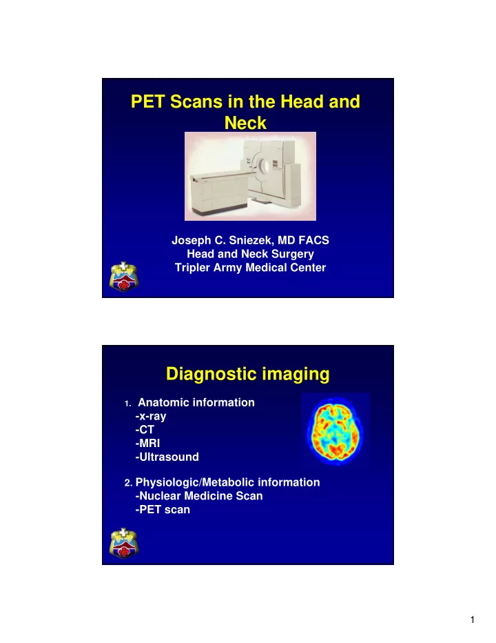
Diagnostic imaging
- 1. Anatomic information
- x-ray
- CT
- MRI
- Ultrasound
- 2. Physiologic/Metabolic information
- Nuclear Medicine Scan
- PET scan