Distinctive Lymphomas Head & Neck Lymphomas: Update on - - PowerPoint PPT Presentation

distinctive lymphomas
SMART_READER_LITE
LIVE PREVIEW

Distinctive Lymphomas Head & Neck Lymphomas: Update on - - PowerPoint PPT Presentation

Lymphomas of the Head and Neck Natkunam & Warnke (2011) 12 Patrick Treseler, MD, PhD University of California San Francisco Distinctive Lymphomas Head & Neck Lymphomas: Update on Distinctive Entities of the Head & Neck 1.


slide-1
SLIDE 1

1

Lymphomas of the Head and Neck

Patrick Treseler, MD, PhD University of California San Francisco

Natkunam & Warnke (2011)12

Head & Neck Lymphomas: Update on Distinctive Entities

  • 1. Extranodal marginal zone lymphoma
  • f mucosa-associated lymphoid tissue

(MALT lymphoma)

  • 2. Extranodal NK/T-cell lymphoma,

nasal type

  • 3. Plasmablastic lymphoma

Distinctive Lymphomas

  • f the Head & Neck
  • 1. Extranodal marginal zone lymphoma
  • f mucosa-associated lymphoid tissue

(MALT lymphoma)

  • 2. Extranodal NK/T-cell lymphoma,

nasal type

  • 3. Plasmablastic lymphoma
slide-2
SLIDE 2

2

Clinical features:

  • Typically arises in setting of lymphoid hyperplasia

associated with autoimmune disorders (LESA, Hashimoto’s thyroiditis) or chronic infection (conjunctivitis due to C. psittaci)

  • Can present as mass/swelling or no symptoms
  • Most low stage (I/II) at presentation, good prog.

MALT lymphoma of H&N

P.G. Isaacson & A.J. Norton. Extranodal Lymphomas. New York: Churchill Livingstone,1994

Neoplastic Small Lymphocytes in MALT Lymphoma

“Resemble centrocytes” “Resemble small lymphocytes” “Resemble monocytoid B-cells”

slide-3
SLIDE 3

3

Immunophenotype:

  • Most have “B-cell, NOS” phenotype, but aberrant

CD43 expression seen in nearly half of cases

  • Plasma cells monotypic in about one-third of cases
  • CD5+ cases rare but well described

MALT lymphoma of H&N

CD20 CD43

slide-4
SLIDE 4

4

Benign:

  • Lymphoepithelial sialadenitis (LESA)
  • Chronic sclerosing sialadenitis

MALT lymphoma of H&N

Malignant:

  • Lymphoplasmacytic lymphoma and
  • ther small B-cell lymphomas
  • Extramedullary plasmacytoma

Differential Diagnosis Benign:

  • Lymphoepithelial sialadenitis (LESA)
  • Chronic sclerosing sialadenitis

MALT lymphoma of H&N

Malignant:

  • Lymphoplasmacytic lymphoma and
  • ther small B-cell lymphomas
  • Extramedullary plasmacytoma

Differential Diagnosis

Lymphoepithelial sialadenitis

Previously known as myoepithelial sialadenitis (MESA), benign lymphoepithelial lesion, & Mikulicz disease Characterized by marked infiltration of small lymphocytes (mainly T-cells) into atrophic salivary gland parenchyma May form mass or be asymptomatic LESA increase risk of salivary gland lymphoma up to 44-fold (most are MALT lymphomas)4

slide-5
SLIDE 5

5

CD20

Lymphoepithelial sialadenitis

Transformation to MALT lymphoma

Monocytoid small B-cells not only in the LELs, but

  • utside as well, forming broad halos or sheets

Sheets of plasma cells (stain for kappa/lambda)

Earliest histopathologic indicators:

slide-6
SLIDE 6

6

CD20

slide-7
SLIDE 7

7

Benign:

  • Lymphoepithelial sialadenitis (LESA)
  • Chronic sclerosing sialadenitis

MALT lymphoma of H&N

Malignant:

  • Lymphoplasmacytic lymphoma and
  • ther small B-cell lymphomas
  • Extramedullary plasmacytoma

Differential Diagnosis

Chronic sclerosing sialadenitis

Also known as Küttner tumor, and almost exclusively affects submandibular gland, producing large firm gland that mimics tumor One of the family of IgG4-related fibrosclerosing diseases that include autoimmune pancreatitis, but has been described in virtually every organ Major histopathologic features are 1) dense lymphoplasmacytic infiltrate, 2) storiform fibrosis, and 3) obliterative phlebitis, but latter two features can be absent in head & neck sites (salivary glands, lacrimal gland)! 3

Deshpande et al. Mod Pathol 25: 1181; 2012.

IgG4-Related Disease of Salivary Gland

Deshpande et al. (2012) Mod Pathol 25:1181.

slide-8
SLIDE 8

8

Benign:

  • Lymphoepithelial sialadenitis (LESA)
  • Chronic sclerosing sialadenitis

MALT lymphoma of H&N

Malignant:

  • Lymphoplasmacytic lymphoma and
  • ther small B-cell lymphomas
  • Extramedullary plasmacytoma

Differential Diagnosis

Small B-Cell Lymphomas

Basic Immunophenotypes

CD20 CD5 CD43 CD23 BCL1 BCL6 CD10

CLL/SLL + + + +

  • Mantle cell

+ + +

  • +
  • Follicular

+

  • /+
  • +

+/- MZL/LPL +

  • /+
  • Proportion of cases positive: + >90%, +/- 50-90%, -/+ 10-50%, - <10%

(Cyclin D1)

Benign:

  • Lymphoepithelial sialadenitis (LESA)
  • Chronic sclerosing sialadenitis

MALT lymphoma of H&N

Malignant:

  • Lymphoplasmacytic lymphoma and
  • ther small B-cell lymphomas
  • Extramedullary plasmacytoma

Differential Diagnosis

slide-9
SLIDE 9

9

Extramedullary Plasmacytoma

Clonal tumor-forming neoplasm of plasma cells

  • ccurring outside the bone/bone marrow in the

absence of a clinical evidence of myeloma at presentation 80% occur in upper aerodigestive tract, most commonly in nasal cavity, paranasal sinuses, nasopharynx, & oropharynx, usually good prognosis Composed of sheets of mature plasma cells, with typical plasma cell phenotype (CD20- CD138+ cIg+) But extranodal MZL can have variable proportions of mature plasma cells, occ. quite high, causing them to resemble EMP

Kappa Lambda CD20

slide-10
SLIDE 10

10

Question Time!

What is the difference between an extranodal marginal zone lymphoma with extensive/extreme plasmacytic differentiation and a plasmacytoma? Maybe nothing (at least in some cases) Q: A:

Am J Clin Pathol 111: 111; 1995

Extramedullary Plasmacytoma: A Form of Extranodal MZL?

Hussong et al. AJCP 111:111; 1995

Predilection for mucosa surfaces Often have some monocytoid or CCL cells Often form LELs if epithelium present Some have classic extranodal MZL before or after dx EMP Do well with conservative therapy, no progression to myeloma

Extra-osseous plasmacytomas can share many features with MALT lymphoma:

“We hypothesize that EMPs not associated with MM, whether in mucosal sites or LNs, may represent MZL with extensive plasmacytic differentition”

Plasmacytoid Marginal Zone Lymphoma vs. Plasmacytoma A not unreasonable approach:

Regard extraosseous tumors of mature plasma cells as MZL or MALT lymphoma with extensive

  • r extreme plasmacytic differentiation if they:

Present in sites commonly involved by

MALT lymphoma

Form lymphoepithelial lesions Don’t aberrantly express CD56 or BCL-1

(common in myeloma, uncommon in EMP)

Clinical work-up for myeloma is negative!

slide-11
SLIDE 11

11

Distinctive Lymphomas

  • f the Head & Neck
  • 1. Extranodal marginal zone lymphoma
  • f mucosa-associated lymphoid tissue

(MALT lymphoma)

  • 2. Extranodal NK/T-cell lymphoma,

nasal type

  • 3. Plasmablastic lymphoma

Extranodal NK/T-cell Lymphoma, Nasal Type

Key Features:

Malignant lymphoma that typically co-expresses:

  • NK-cell marker CD56, T-cell markers (CD2, CD43, CD45RO,

and sometimes CD7), & cytotoxic proteins (TIA-1, etc.)

  • But usually not CD5, CD4, CD8, TCRαβ, or TCRγδ.

Surface CD3- (flow), but cytoplasmic CD3ε+ (IHC) Almost universally EBV+ by ISH (“diagnosis should be accepted with some skepticism if EBV is negative”) Most are NK-cells (TCR germline), minority are T-cells (TCR rearranged), but behavior & treatment are similar Nasal cavity, nasopharynx, palate most common sites Cytologic composition highly variable, often angiocentric

“Settled Law” in the DDx

  • f NK/T-Cell Lymphoma

(per 2008 WHO)

If a CD3ε+ lymphoma is CD56 negative, it can still be NK/T-cell lymphoma, provided it is positive for both:

  • EBV (i.e., EBER), and
  • Cytotoxic proteins (i.e., TIA-1, granzyme B, perforin)

But if negative for either EBV or cytotoxic granule proteins (while still CD3ε+), it goes to category of PTCL, NOS Remember, many of these lymphomas are clonal T-cell neoplasms, and they can lack NK marker CD56, but still need to be cytotoxic T-cells (and probably EBV+ too)!

slide-12
SLIDE 12

12

CD3

slide-13
SLIDE 13

13

CD5 CD56 EBV-ISH

slide-14
SLIDE 14

14

CD56

Question Time!

Can you have an extranasal extranodal nasal-type NK/T-cell lymphoma? Yes, well described in skin, GI tract, lung, testis, eye, other sites per WHO. Q: A:

Question Time!

Are there NK/T-cell lymphomas

  • ther than nasal-type?

Apparently not. NK-cell neoplasms in 2008 WHO are:

  • Extranodal NK/T-cell lymphoma, nasal type
  • Aggressive NK-cell leukemia
  • Chronic lymphoproliferative disorder of NK cells
  • NK lymphoblastic leukemia/lymphoma

Q: A:

Question Time!

Can you have a nodal extranodal nasal-type NK/T-cell lymphoma? Yes, 2008 WHO cites rare reports of primary lymph node disease in the absence of extranodal disease (but “extranodal” part of official WHO name) Q: A:

slide-15
SLIDE 15

15

NK/T-cell Lymphoma

Differential Diagnosis:

Benign:

  • Reactive lymphoid hyperplasia

Malignant:

  • Peripheral T-cell lymphoma, NOS
  • Other NK-cell malignancies
  • Other CD56+ malignancies

NK/T-cell Lymphoma

Differential Diagnosis:

Benign:

  • Reactive lymphoid hyperplasia

Malignant:

  • Peripheral T-cell lymphoma, NOS
  • Other NK-cell malignancies
  • Other CD56+ malignancies

Reactive Lymphoid Hyperplasia

Mimics NK/T-cell lymphoma because:

  • Extranodal reactive lymphoid cells often mildly

atypical, form dense and monomorphic infiltrates that are mixture of T-cells and NK-cells with few B-cells

  • In Wegener’s, may be angiocentric with necrosis

Not NK/T-cell lymphoma because:

– Reactive hyperplasia will not show sheet-like growth

  • f CD3ε+ CD56+ TIA-1+ EBER+ cells

– Typically lacks destructive infiltration of mucosa, ulceration, high mitotic rate often seen in NK/TCL – Never show high-grade cytologic atypia of virtually all cells seen in many cases of NK/TCL

NK/T-cell Lymphoma

Differential Diagnosis:

Benign:

  • Reactive lymphoid hyperplasia

Malignant:

  • Peripheral T-cell lymphoma, NOS
  • Other NK-cell malignancies
  • Other CD56+ malignancies
slide-16
SLIDE 16

16

Peripheral T-cell lymphoma

Lack of CD56 and both EBV and cytotoxic proteins puts them in category of PTCL, NOS per current WHO criteria “Gray zone” could exist for cases showing expression of multiple T-cell markers not typical of NK/TCL (CD5, CD4, CD8, TCRαβ, and/or TCRγδ), particularly if EBV negative PTCL, NOS can show variable expression of CD56, EBV, and cytotoxic proteins, but in isolation these are insufficient for NK/TCL dx

NK/T-cell Lymphoma

Differential Diagnosis:

Benign:

  • Reactive lymphoid hyperplasia

Malignant:

  • Peripheral T-cell lymphoma, NOS
  • Other NK-cell malignancies
  • Other CD56+ malignancies

Other NK-cell malignancies

WHO recognizes three such disorders, all rare:

− Aggressive NK-cell leukemia (EBV+) − Chronic lymphoproliferative disorder of NK-cells − NK-lymphoblastic leukemia/lymphoma

Tissue infiltrates could mimic NK/T-cell lymphoma because of NK-cell phenotype All distinguishable from NK/T-cell lymphoma

  • n basis clinical features (presentation as

aggressive leukemia) and/or phenotype (weak to absent CD56, absent EBV)

NK/T-cell Lymphoma

Differential Diagnosis:

Benign:

  • Reactive lymphoid hyperplasia

Malignant:

  • Peripheral T-cell lymphoma
  • Other NK-cell malignancies
  • Other CD56+ malignancies
slide-17
SLIDE 17

17

Other CD56+ malignancies

Many high-grade malignancies can appear vaguely lymphoid and express CD56, sometimes along T-cell markers, and arise in head and neck, principally:

− Myeloid sarcoma/acute myeloid leukemia − Blastic plasmacytoid dendritic cell neoplasm − Plasmacytoma/myeloma − Ewing sarcoma/PNET − Rhabdomyosarcoma

All distinguishable from NK/T-cell lymphoma

  • n basis clinical features and/or phenotype

Distinctive Lymphomas

  • f the Head & Neck
  • 1. Extranodal marginal zone lymphoma
  • f mucosa-associated lymphoid tissue

(MALT lymphoma)

  • 2. Extranodal NK/T-cell lymphoma,

nasal type

  • 3. Plasmablastic lymphoma

Plasmablastic Lymphoma

Aggressive variant of diffuse large B-cell lymphoma Composed of large cells +/- plasmacytoid differentiation CD20 and Pax-5 expression generally negative but weak staining (<25% of cells) in some cases, CD45- or weak Often presents in oral cavity, but can also involve other mainly extranodal sites Patients mainly HIV+, other immunocompromised Frequently EMA+, rare keratin+ cases reported Very poor prognosis, median survival 6-7 months

Key Features:

CD34 CD19 CD20 Pax-5 Surface Ig CD10 CD10 CD5 CD138 BOB.1

Germinal Center

Lymphoblastic leukemia/lymphoma CLL/SLL Mantle cell DLBCL? Follicular lymphoma Burkitt lymphoma DLBCL MZL DLBCL Plasmacytoma Plasmablastic lymphoma

Ig heavy chain genes rearranged Ig light chain genes rearranged Ig genes hypermutated

CD79a Oct-2

slide-18
SLIDE 18

18

Pax-5 CD20 MUM1

slide-19
SLIDE 19

19

Oct-2

Plasmablastic Lymphoma

Plasmablastic lymphoma,

  • ral mucosa type

Plasmablastic lymphoma with plasmacytic differentiation

(AJSP 28:736; 2004)

Subtypes per Colomo et al.

PBL, oral mucosa type

PBL, oral mucosa type. Colomo et al. AJSP 28:736.

slide-20
SLIDE 20

20

PBL, with plasmacytic diff.

PBL with plasmacytic differentiation. Colomo et al. AJSP 28:736. Secondary Extramedullary Plasmablastic Tumor (EPT)

Colomo et al. AJSP 28:736.

slide-21
SLIDE 21

21

Plasmablastic lymphoma

Differential Diagnosis:

Malignant:

  • Anaplastic plasmacytoma
  • ALK+ diffuse large B-cell lymphoma
  • Poorly differentiated non-lymphoid

malignancies, esp. undifferentiated nasopharyngeal ca, Schmincke pattern

Plasmablastic lymphoma

Differential Diagnosis:

Malignant:

  • Anaplastic plasmacytoma*
  • ALK+ diffuse large B-cell lymphoma
  • Poorly differentiated non-lymphoid

malignancies, esp. undifferentiated nasopharyngeal ca, Schmincke pattern

* Original term used for tumor that would today be called PBL!14

Plasmablastic lymphoma

Differential Diagnosis:

Malignant:

  • Anaplastic plasmacytoma
  • ALK+ diffuse large B-cell lymphoma
  • Poorly differentiated non-lymphoid

malignancies, esp. undifferentiated nasopharyngeal ca, Schmincke pattern

ALK+ large B-cell lymphoma

Very rare variant of DLBCL (<40 cases reported) Wide age range pediatric → adult Typically presents in lymph nodes, but some extranodal (including nasopharynx, tongue), often advanced stage Tumor cells resemble immunoblasts or plasmablasts, but can be RS-like, and typically infiltrate lymph node sinuses Has CD20- Pax5- Oct2+ plasma cell phenotype, CD45- or weak (overlap with plasmablastic lymphoma). Some cases EMA+ and keratin+ ALK staining typically cytoplasmic & granular, and due to t(2;17) linking ALK with CLTC gene. Median surv. 11 mo. (some pediatric long-term survivors)

slide-22
SLIDE 22

22

AJSP 29: 1652; 2005

P = 0.03 P = 0.003 P = 0.008 P = 0.32 (NS)

Laurent, C. et al. J Clin Oncol; 27:4211-4216; 2009

ALK

Plasmablastic lymphoma

Differential Diagnosis:

Malignant:

  • Anaplastic plasmacytoma
  • ALK+ diffuse large B-cell lymphoma
  • Poorly differentiated non-lymphoid

malignancies, esp. undifferentiated nasopharyngeal ca, Schmincke pattern

UNPC, Schmincke Pattern

UNPC is a subtype of non-keratinizing nasopharyngeal carcinoma in 2005 WHO Classification Typically small primary with cervical lymph LN metastases Tumor cells large cells and loosely cohesive (“syncytial growth pattern”) mimicking large lymphoid cells, with heavy background infiltrate of small lymphocytes In Schmincke pattern, tumor cells present as single cells, small clusters, or ill-defined sheets, mimicking lymphoma (Regaud pattern has more obvious epithelial aggregates and sheets). Tumor cells EBV+, up to 20% keratin-negative, creating further confusion with large cell lymphomas such as PBL

slide-23
SLIDE 23

23

EBV-ISH Pan-CK

References

  • 1. Chan ACL, Chan JKC. Hematolymphoid lesions of the head and neck. In: Surgical

Pathology of the Head and Neck, 3rd ed. (L. Barnes, ed). New York: Informa Healthcare USA, 2009.

  • 2. Colomo L, et al. Diffuse large B-cell lymphomas with plasmablastic differentiation

represent a heterogeneous group of disease entities. Am J Surg Pathol. 2004 Jun;28(6): 736-47.

  • 3. Deshpande V, et al. Consensus statement on the pathology of IgG4-related
  • disease. Mod Pathol. 2012 Sep;25(9):1181-92.
  • 4. Eveson JW, Nagao T. Diseases of the salivary glands. In: Surgical Pathology of the

Head and Neck, 3rd ed. (L. Barnes, ed). New York: Informa Healthcare USA, 2009.

  • 5. Franchi A, et al. Sinonasal undifferentiated carcinoma, nasopharyngeal-type

undifferentiated carcinoma, and keratinizing and nonkeratinizing squamous cell carcinoma express different cytokeratin patterns. Am J Surg Pathol. 2002 Dec; 26(12): 1597-604.

  • 6. Hussong JW, et al. Extramedullary plasmacytoma. A form of marginal zone cell

lymphoma? Am J Clin Pathol. 1999 Jan; 111(1): 111-6.

  • 7. Isaacson PG, Norton AJ. Extranodal Lymphomas. New York: Churchill Livingstone,

1994.

References

  • 8. Isaacson PG, et al. Extranodal marginal zone lymphoma of mucosa-associated

lymphoid tissue. In: WHO Classification of Tumours of Haematopoietic and Lymphoid Tissues (Swerdlow et al, eds.). Lyon, France: International Agency for Research on Cancer, 2008.

  • 9. Jaffe ES, et al. Hematopathology. Philadelphia: Saunders/Elsevier, 2011.

10.Kim JE, et al. Human immunodeficiency virus-negative plasmablastic lymphoma in

  • Korea. Leuk Lymphoma. 2009 Apr;50(4): 582-7.

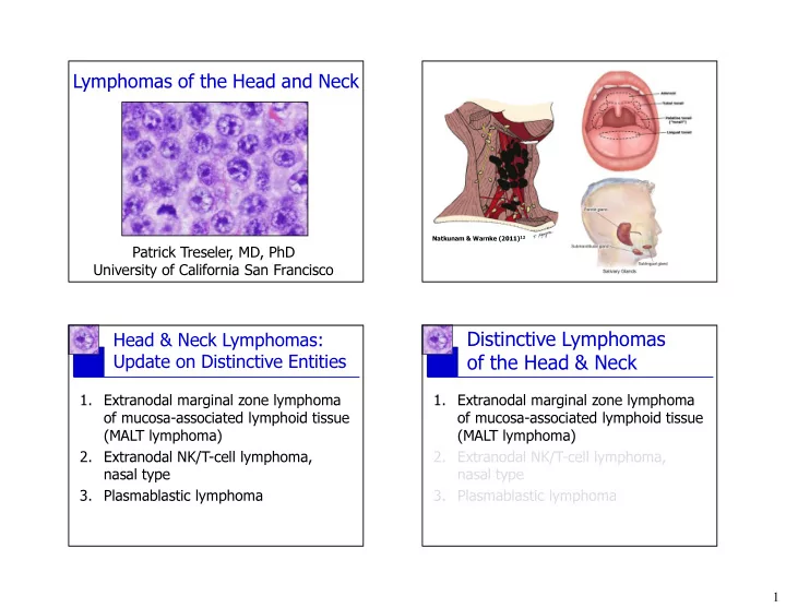
11.McKenna RW, et al. In: WHO Classification of Tumours of Haematopoietic and Lymphoid Tissues (Swerdlow et al, eds.). Lyon, France: International Agency for Research on Cancer, 2008. 12.Natkunam Y, Warnke RA. Processing of the lymph node biopsy specimen. In: Hematopathology (Jaffe at al, eds). Philadelphia: Saunders/Elsevier, 2011. 13.Nomori H, et al. Nasopharyngeal carcinoma: immunohistochemical study for keratin, secretory component and leukocyte common antigen. Jpn J Clin Oncol. 1985 Mar; 15(1): 95-105. 14.Sperling RI, Fromowitz FB, Castellano TJ. Anaplastic solitary extramedullary plasmacytoma of the cecum. Report of a case confirmed by immunoperoxidase

  • staining. Dis Colon Rectum. 1987 Nov; 30(11): 894-8.
slide-24
SLIDE 24

24

References

15.Swerdlow SH, et al. WHO Classification of Tumours of Haematopoietic and Lymphoid Tissues. Lyon, France: International Agency for Research on Cancer, 2008.

CD20 Pan-CK

slide-25
SLIDE 25

25

CD3

Kappa CD20

slide-26
SLIDE 26

26

EMA CD3 CD20