Overview Introduction Evaluation Imaging Management SURGICAL - - PowerPoint PPT Presentation

overview
SMART_READER_LITE
LIVE PREVIEW

Overview Introduction Evaluation Imaging Management SURGICAL - - PowerPoint PPT Presentation

HCC Overview Introduction Evaluation Imaging Management SURGICAL MANAGEMENT Supportive Care HEPATOCELLULAR CANCER Palliative Carlos U. Corvera M.D. Associate Professor Curative Department of Surgery Chief,


slide-1
SLIDE 1

1

Carlos U. Corvera M.D. Associate Professor Department of Surgery Chief, Liver, Biliary and Pancreatic Surgery University of California, San Francisco School of Medicine

SURGICAL MANAGEMENT HEPATOCELLULAR CANCER

UCSF 2013 Postgraduate Course HCC

Overview

  • Introduction
  • Evaluation
  • Imaging
  • Management

– Supportive Care – Palliative – Curative

HCC

Primary Liver Cancer

  • 5th leading cause of cancer in the world
  • Heterogeneous incidence

Low in America and Northern Europe Intermediate in South Europe High in Subsaharian Africa and Far East

  • In the U.S. > 20,000 new cases in 2000
  • 75% increase since 1993
  • Rising incidence of chronic hepatitis in U.S.
  • 1.2 million cases of hepatitis B
  • 3 million cases of hepatitis C
  • Predicted to equal levels in Japan within two decades

NEJM 1999; 340.745 PNAS 2002;99:15584-89

HCC

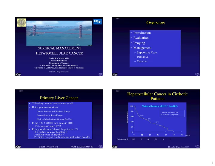
Hepatocellular Cancer in Cirrhotic Patients

20 40 60 80 100 12 24 36 48 60 72 Probability (%)

months

Patients at risk 102 57 40 21 8 1

Median surviva l=17m # of deaths = 79 patients 54% 40% 28% Llovet JM. Hepatology, 1999.

Natural history of HCC (n=102)

slide-2
SLIDE 2

2

HCC

HCC Carcinogenic Sequence

  • Solitary- encapsulated
  • Multifocal /multinodular,

Bilobar

  • Diffusely Infiltrative/ Invasive
  • Regenerative Nodule

(size criteria)

  • Dysplastic Nodule

Dysplasia Neoplasia

3- 50 months

Early HCC

HCC

Imaging of the Liver

  • Ultrasound

– Contrast

  • Computed

Tomography

– Multi Detector Technology

  • Magnetic Resonance

– Ultrafast Imaging – Diffusion Weighted – Eovist

  • Lesion characterization
  • Lesion detection
  • Staging for potential

resection

  • Assessment of

therapeutic response GOALS OPTIONS

HCC

Hepatocellular Carcinoma

  • Receives its blood supply from the hepatic artery and

consists of abnormal hepatocytes arranged in a trabecular, sinusoidal pattern

  • Expansive tumors: well differentiated and relatively slowly

growing; usually well defined

  • Invasive tumors: poorly differentiated with aggressive

growth patterns; usually ill defined

  • Invades vascular structures, more commonly the portal vein

than the hepatic vein; arterioportal shunting is characteristic

HCC

Duplex Ultrasound Left Hepatic Vein Tumor Involvement

trans left

HCC

slide-3
SLIDE 3

3

HCC

Portal Vein Thrombosis

Long Long Trans Trans

HCC

Hepatocellular Carcinoma

trans MHV RHV IVC long

HCC

Computed Tomography

  • Spiral
  • Multidetector Spiral CT
  • Major Advances

– 3D Reformatting – CT Angiography

  • Hyperdense enhancement

during arterial phase

  • Lesion become lower in

density during later phase i.e “Washout”

Advances HCC Characteristics on CT

HCC

Multiphasic CT-Scan

Example 1 Example 2

slide-4
SLIDE 4

4

HCC

MR Imaging

  • Conventional Imaging

– T1 (anatomy) – T2 (pathology) – Flow (MR angiography)

  • Others

– Fat (lesion characterization) – Metabolites (spectroscopy) – Tissue oxygen consumption (fMRI) – Diffusion & perfusion (ischemia, necrosis) – Temperature (monitoring therapy)

HCC

HCC

MRI

HCC

Gadolinium Arterial Delayed 5 min

MR Imaging

Gadolinium Portal Venous Delayed 1 min Coronal Pre-Gadolinium

HCC

  • Supportive Care
  • Palliative Therapies

– Transarterial embolization (TAE) or Chemoembolization (TACE) – Percutenous Ablative Procedures – Hormonal treatments/ Immunotherapy – Antiproliferative agents – Radiation Therapy: external and transarterial

  • Curative (Radical) Therapies

– Surgical resection (5-40%) – Liver Transplantation (CLT/LDLT) – Ablative procedures : Percutaneous ethanol injection (PEI) /Radiofrequency

Management of HCC

Multidisciplinary: Hepatologist, Oncologist, Diagnostic Radiologist,

Interventional Radiologist and Surgeons

slide-5
SLIDE 5

5

HCC

Ablation Tools

  • Chemical

– ETOH – Acetic Acid – Chemotherapy – Experimental protocols

  • Thermal

– RF ablation – Cryoablation – Laser – Microwave

HCC

Percutaneous Ethanol Injection (PEI)

  • Need to be able to locate the lesion
  • Should be < 3cm

HCC

PEI 6 months later

HCC

Principles of Embolization Therapy

  • Dual blood supply to liver facilitates preferential

delivery of embolic/toxic agents to tumor “sparing” normal liver

  • Access to hepatic artery allows for targeted

regional therapy, minimal systemic effect

slide-6
SLIDE 6

6

HCC

Patient Selection for TAE or TACE

  • Unresectable disease

– Multiple small tumors – Large >5 cm involving critical structures

  • Liver only/dominant disease
  • Adequate hepatic functional reserve

Labs: T.bili< 2.0; Cr. < 1.5, Plts> 75

  • Most are palliative procedures-

HCC

Tumor

EARLY ARTERIAL

CT Scan

NON-CONTRAST

Tumor

HCC

Single Lesion Embolization

Celiac -- Scout

Tumor

HCC

Single Lesion Embolization

Left HA Selective Left HA Post-embolization Left HA

Tumor Tumor

slide-7
SLIDE 7

7

HCC

Multiple / Bilobar Disease

HCC

Left-sided tumors

Multiple / Bilobar Disease

HCC

Embolization Procedures

  • -Complications--
  • Occlusion of vessels to non-target organs

– cystic artery chemical cholecystitis – right gastric artery gastric or duodenal ulceration – GDA Acute pancreatitis – Biliary Necrosis (dilated intrahepatic ducts)

  • Catheter related vascular injuries

– Hemorrhage – Dissections – Aneurysms – Puncture site hematoma

  • Liver Abscess
  • Liver Decompensation
  • Post-Embolization Syndrome

HCC

Patient Selection for Thermal Ablations

  • Non-operative potential --quality of life issues.
  • Unresectable
  • Anatomy – often not good for RFA either
  • Extending limits of resection
  • Tumor Characteristics: Size and Distribution

– Number < 3 – Size < 3cm , < 5 cm

  • Assuming perfect accuracy should be as good as

resection

slide-8
SLIDE 8

8

HCC

Thermal Ablation – Guidance

  • Ultrasound
  • Computed Tomography
  • Magnetic Resonance Imaging

HCC

RFA/Microwave – Techniques

  • Percutaneous: CT scan/ US
  • General anesthesia not always necessary
  • Less trauma/pain/recovery
  • Some tumors not anatomically feasible
  • No operative staging
  • Laparoscopic /Thoracoscopic
  • Minimally invasive surgery
  • Better staging
  • Able to move organs from heat source
  • Open
  • Optimal staging
  • ? Optimal probe placement
  • Able to combine with resection

HCC

CT – Guidance

HCC

Laparoscopic Technique

Umbilical Vein

slide-9
SLIDE 9

9

HCC HCC HCC

RFA/Microwave – Local failure

  • Increasing size
  • Tumor vascularity
  • Proximity to vascular structures
  • Surgical versus percutaneous technique

HCC

Preoperative MRI

slide-10
SLIDE 10

10

HCC

Post-RFA MRI

6 months 14 months

HCC

Curative Therapies

  • Surgical Resection
  • Liver Transplantation
  • Ablative Therapies

HCC

Two Problems

  • Chronic Liver Disease & HCC
  • Underlying cirrhosis limits

aggressive treatments.

  • Surgery remains the only chance

for long-term survival.

  • Majority of patients are not

suitable for operation.

Tumor

HCC

“…Partial hepatectomy for tumors occurring in cirrhotic livers should not be done unless it is necessary to control hemorrhage.”

  • Liver Tumor Survey-- 1974
  • Mortality rate was 58% in cirrhotic patients

(n =26).

Foster JM, Berman MM,. Solid Liver Tumor,1977;p. 62-104

Hepatic Resection in Cirrhotic Livers: The Early View

slide-11
SLIDE 11

11

HCC

Preoperative Assessment of Liver Function

*alone not considered an absolute contraindication to resection

Test Author Contraindication for Resection

Child-Pugh Franco Score > 8 Serum alanine Noun ALT > twofold upper limit of norm Indocyanine green Lau retention rate at 15 minutes > 15 % Makuuchi retention rate at 15 minutes > 10 % Fan retention rate at 15 minutes > 14 % Wu retention rate at 15 minutes > 10 % Hasegawa retention rate at 15 minutes > 10 % Hemming clearance < 5 mL/min/kg Kanematsu retention rate at 15 minutes > 20 % Urea nitrogen synthesis Paquet < 6 g/day Portal Vein Pressure Bruix HVPG > 10mm Hg Lidocaine (MEGX) test Ercolani G MEGX <25 ng/ml* Grazi, MEGX <25 ng/ml*

HCC

Preoperative Interventions

  • Prevention of Variceal Bleeding

– Sclerotherapy – Transjugular intrahepatic portosystemic shunt (TIPS)

  • Arterial Embolization

– Diagnostic Angiogram – Reduces tumor bulk

  • Sequential Arterial and Portal

Embolization (double vascular embolization).

Varices Umbilical Vein

HCC

Right Hepatic Artery Embolization

Replaced Right Hepatic Artery Post embolization

Sequential Arterial and Portal Embolization

HCC

3 Weeks After Right Hepatic Arterial Embolization

slide-12
SLIDE 12

12

HCC

Right Portal Vein Embolization

Occluded PV Occluded PV

Sequential Arterial and Portal Embolization

HCC

Right Portal Vein Embolization

Occluded PV Occluded PV

HCC

Repeat Right & Left Hepatic Arterial Embolization

Recanulated Branch from Replaced RHA Left HA Branches

HCC

CT Scan

6 weeks after PVE, 9 & 3 weeks after TACE

slide-13
SLIDE 13

13

HCC

Operation

HCC

Postoperative CT Scan

HCC

General Operative Considerations

  • Specific technical difficulties in the Cirrhotic:

– Parenchyma is hard. – Anatomic landmarks are distorted. – Tissue friability. – Tumors may be difficult to recognized from the surrounding cirrhotic liver.

*Yoshida Y, Ann of Surg 1989;209 (3):297-301

HCC

  • Complete Staging: Bone scan, CT-chest
  • Preserved Liver Function
  • Childs A-- ONLY
  • Cardiopulmonary assessment (> 65 yrs).
  • Open Resections are now rare: Majority are done

Laparoscopically, or Not at all.

  • When done open, we prefer a Makuuchi incision
  • Full abdominal exploration.
  • Intraoperative ultrasound.

General Operative Considerations

slide-14
SLIDE 14

14

HCC

Non-Anatomic Resections

HCC

Operation

HCCOperative Management of Large HCC

  • -Anterior Approach--

HCC

Large HCC –Anterior Approach

slide-15
SLIDE 15

15

HCC

L Hepatectomy L Extended Hepatectomy R Extended Hepatectomy R Hepatectomy

Extended Resections

HCC

Extended Resections

HCC

Extended Resections

HCC

Combining Modalities

  • Most patients are treated by multiple

complementary ablative approaches

  • The order depends on the pattern of disease
  • Options include

– Resection, RFA/microwave, TACE, and/or PEI – RFA/microwave then TACE – PEI then, TACE

slide-16
SLIDE 16

16

HCC

Tumor Invading R Kidney

Presentation CT Scan

HCC

Venous Shunt Renal Pelvis

Hepatic Arterial Embolization

Right (posterior) HA Venous Shunt

HCC

Right Renal Artery Embolization

HCC

Post Embolization Right posterior HA & Superior Pole Renal Artery

slide-17
SLIDE 17

17

HCC

Repeat RHA (entire) Embolization

HCC

Residual Tumor

CT Scan Following TAE x 5

HCC

CT Scan Following TAE x 6

Enhancing tumor

HCC

Percutaneous Ultrasound Guided RFA of Residual Liver Tumor

Kidney

slide-18
SLIDE 18

18

HCC

Post RFA Pre RFA

HCC

AFP levels

RFA TAE

HCC

Multiple Modality Approach

HCC

Post Laparoscopic Resection and RFA

slide-19
SLIDE 19

19

HCC

CT Scan 12 months later TAE x 3 Post Treatment CT Scan (3 months)

PEI & TAE Enlarged Lesion

HCC HCC

Summary

  • Therapy of liver tumors in patients with cirrhosis

remains challenging.

  • However, hepatic resection for HCC can be

performed safely, provided a limited resection is anticipated.

  • Resection should be considered the standard

therapy for HCC.

  • Inoperable patients can benefit from liver

directed transarterial ablative therapies to halt tumor progression and to extend survival.

CONTACT INFORMATION: Carlos U. Corvera M.D. Associate Professor Department of Surgery Gastrointestinal Surgical Oncology Chief, Liver, Biliary and Pancreatic Surgery University of California, San Francisco School of Medicine Helen Diller Family Comprehensive Cancer Center Email: carlos.corvera@ucsfmedctr.org Phone: Mobile # (415) 317-4602 Direct Line: (415) 502-1690 Clinic Line (referrals) Fax: (415) 353-9931 Tel: (415) 353-9888 Academic Practice: Address: Room U-370 521 Parnassus Avenue San Francisco, CA. 94143 Assistant (Marjorie Galicha) email: Marjorie.Galicha@ucsfmedctr.org tel: (415) 415-476-0762