
SLIDE 1
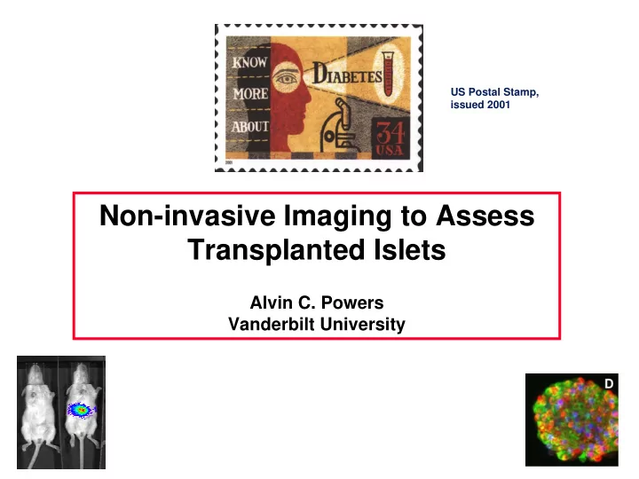
US Postal Stamp, issued 2001
Non-invasive Imaging to Assess Transplanted Islets
Alvin C. Powers Vanderbilt University
Non-invasive Imaging to Assess Transplanted Islets Alvin C. Powers - - PowerPoint PPT Presentation
US Postal Stamp, issued 2001 Non-invasive Imaging to Assess Transplanted Islets Alvin C. Powers Vanderbilt University Today Rationale and challenges for imaging pancreatic islets Overview of imaging modalities being used
US Postal Stamp, issued 2001
Alvin C. Powers Vanderbilt University
Rationale and challenges for imaging
Overview of imaging modalities being used Bioluminescence to assess transplanted
Human Islet
α cells β cells δ cells
Pancreas
Purified Human Islets
Photo courtesy of David Harlan Photo courtesy of David Harlan
Islets size ( < 250 µm) is less than resolution of imaging modalities (CT, MRI)
Transplanted, intra-hepatic islets are
Techniques to non-invasively assess or
Difficult to study islet survival following
Cannot assess interventions to sustain or
Non-invasive and allow for serial, in vivo
Non-toxic to islet cells Useful for study of islets in native pancreas
Applicable to murine models Adaptable for human imaging
Should it measure islet cell mass or number?
Should it reflect function or health of beta
Is spatial resolution of islets important?
Pancreas
Utilize an islet-specific protein or process Introduce a reporter into islet cells
Unique glucose metabolism
Islet or beta cell-specific (or
High concentration of zinc
K+ SUR
Insulin
Ca++
Ca++
GLUT2 Glucose Glucokinase ATP/ADP Metabolism
Glucose
Unique glucose metabolism
Islet or beta cell-specific (or
High concentration of zinc In pancreas, islets are highly
Islet in Mouse Pancreas Islet in Mouse Pancreas
MRI PET Bioluminescence Imaging
US Postal Stamp, issued 2001
Luciferase-Luciferin + AMP + O2 Oxyluciferin* + CO2 + AMP Luciferase + Luciferin + ATP + O2 Luciferase-Luciferin + AMP + PPi Oxyluciferin* Oxyluciferin + hν
Luciferase Nucleus Oxygen ATP D-Luciferin Photons
NOD- SCID
Adapted from JDRF figure Adapted from JDRF figure
Accept xenografts Do not develop insulitis or diabetes Allow long-term expression of adenoviral DNA Species-specific insulin assay to distinguish
Lack B- and T-Lymphocytes NOD background further reduces immunity
B.
1000 (no virus) 50
250 500 750 1000 1250 4 8 12 16 r2 = 0.9808 n = 3 wells
# of Islets/well
100 500 1000
1500 Photon counts
500 IEQ 1000 IEQ 2000 IEQ
500 IEQ 1000 IEQ 2000 IEQ
1500 Photon counts 2000 Photon counts
θ θ Camera θ θ Camera
0.2 0.4 0.6 0.8 1 1.2
20 40 60 Angle of Rotation [Degrees] Intensity Normalized to 0 Degrees 0.2 0.4 0.6 0.8 1 1.2
20 40 60 Angle of Rotation [Degrees] Intensity Normalized to 0 Degrees 0.2 0.4 0.6 0.8 1 1.2
20 40 60 Angle of Rotation [Degrees] Intensity Normalized to 0 Degrees 0.2 0.4 0.6 0.8 1 1.2
20 40 60 Angle of Rotation [Degrees] Intensity Normalized to 0 Degrees
400 500 600 700 800 Wavelength [nm] 400 500 600 700 800 Wavelength [nm] 2000 Photon Counts 500 Photon Counts
D E F
0.05 0.1 0.15 0.2 0.25 0.3 1 2 3 4 5 6 7 Weeks Post Implant Ratio of In Vitro Intensity to In Vivo Intensity Renal Hepatic 0.05 0.1 0.15 0.2 0.25 0.3 1 2 3 4 5 6 7 Weeks Post Implant Ratio of In Vitro Intensity to In Vivo Intensity Renal Hepatic
Hepatic Bead Camera Aperture Air 50 cm 5 cm Skin 0.025 cm Liver 0.1 cm Renal Bead
Kidney Liver
50 100 200 20000 40000 60000 80000 100000 120000 Liver Kidney # of Murine Islets Transplanted In vivo bioluminescence (photon counts)
500 1000 1500 2000 2500 4000 8000 12000 16000 20000 r2 = 0.9946 n = 3 - 4 mice 400 800 1200 1600 2000 r2 = 0.9755 n = 3 - 4 mice
Dependent of level of luciferase expression
Optical scattering properties of tissue in which
If these are considered in calculations,
Beneath Renal Capsule
2 4 6 8 10 12 14 16 18 20 1 2 3 4 5
Tx (50 islets)
Weeks post-transplantation In vivo bioluminescence (x10 6) (photon counts)
1 2 3 4 5 6 7 8 9 1 2 3 4 5
Tx (125 islets)
Weeks post-transplantation In vivo bioluminescence (x10 6) (photon counts)
Intrahepatic
Large decline ( > 60%) suggesting islet loss in
Beginning 2 weeks post-transplant, greater loss
Liver is unfriendly site? Destroyed by immune attack against
Islets no longer express luciferase
Cell division and progeny cells no longer
Lentivirus (D. Kaufman, UCLA) AAV virus Transgenic expression of luciferase
Non-invasive Sensitive (detect 25-50 transplanted mouse
Photon generation likely reflects islet cell
Poor spatial resolution Allows for serial measurements of intra-
Allows testing of interventions to increase or
Martin Lepage, John Gore, Vanderbilt Imaging Institute
All imaging modalities will
No single modality will
Understanding islet survival
Marcela Brissova Michael Fowler Wendell Nicholson
Alena Shostak Greg Poffenberger Zhongyi Chen Craig Hauck Jeanelle Kantz Chunhua Dai
Qing Cai Masa Shiota Mark Magnuson Maureen Gannon David Piston John Gore
John Virostko
David Harlan Boaz Hirschberg Mark Atkinson Graeme Bell Soo Young Park Daniel Kaufman