
SLIDE 1
12/12/2015 1
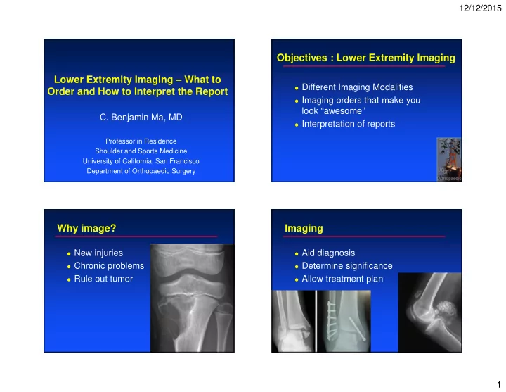
Lower Extremity Imaging – What to Order and How to Interpret the Report
- C. Benjamin Ma, MD
Objectives : Lower Extremity Imaging Lower Extremity Imaging What - - PowerPoint PPT Presentation
12/12/2015 Objectives : Lower Extremity Imaging Lower Extremity Imaging What to Different Imaging Modalities Order and How to Interpret the Report Imaging orders that make you look awesome C. Benjamin Ma, MD Interpretation
Uses high-frequency sound waves to
Similar to sonar wave on getting images of
Can be helpful to evaluate ganglion cyst Knee ganglions Foot ganglions Diagnose tendon tears Foot peroneal tendon injuries Achilles tendon ruptures
Advantages Tomographic evaluation No magnification Give detail in trabecular and cortical
Uses radioisotope-labelled biological
Radioactive tracers administered to
Images produced by scintigraphy Technetium bone scan FDG in PET scans
Rule out tumor – multiple lesions,
Infection – tagged WBC scan Evaluate symptomatic joints Such as arthritis Nonunion Stress fractures
Advantages Imaging of metabolic activity
Diagnosis of infection Disadvantages Lack detail and spatial resolution Limited early sensitivity
Low sensitivity for lytic problems
Helpful to evaluate
Quality of cartilage
fraying arthritis
Labrum and
Helpful to evaluate
Quality of cartilage
fraying arthritis
Labrum and
Helpful to evaluate
Quality of cartilage
fraying arthritis
Labrum and
INDICATION: Age: 17 years. Gender: Male. History:
Bones and joints: Osseous fragment over the superior
Soft tissues: Large joint effusion with patellar soft
IMPRESSION: Osseous fragment over the superior pole of the patella
CLINICAL HISTORY: r/o fx at left 5th MTP.
IMPRESSION:
labrum (image 17, series 4).
ligament are intact. Linear low signal intensity medial to the ligamentum teres may represent a thick acetabular plica.
and iliopsoas tendons are intact. Edema around the gluteus tendon insertion, greater around the minimus than the medius, is compatible with mild peritendinitis.
femoral acetabular cartilage. Focal chondral loss along the posterior medial aspect acetabular cartilage.
around the minimus than the medius.