
SLIDE 1
Mole Mapping: Managing High Risk Patients through Imaging - - PowerPoint PPT Presentation
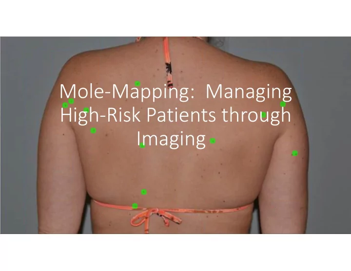
Mole Mapping: Managing High Risk Patients through Imaging Disclosures Chief Medical Officer for MoleSafe USA, LLC Mole Mapping: Managing High Risk Patients through Imaging This session will focus on utilizing imaging
utilizing imaging technology for the management of patients at “high‐risk” for melanoma
A – Asymmetry B – Border Irregularity C – Color Variegation D – Diameter great than 6 mm
E – Evolving
‐ Size ‐ Shape ‐ Symptoms ‐ Surface Bleeding ‐ Shades of Color
Abbasi,N.R.et al. Early Diagnosis of Cutaneous Melanoma.Revisiting The ABCD Criteria .JAMA. 2004;292:2771‐2776
Watts CG, Dieng M, Morton RL, et al. Clinical practice guidelines for identification, screening and follow‐up of individuals at high risk of primary cutaneous melanoma: a systematic review. British Journal of Dermatology. 2015: 33‐47
will increase 7.7%
American Cancer Society – Cancer Facts & Figures 2019 A meta‐analysis of nevus‐associated melanoma: Prevalence and practical implications Pampena, Riccardo et al. Journal of the American Academy of Dermatology, Volume 77, Issue 5, 938 ‐ 945.e4
A recent study published in the JAAD shows that there is general agreement among Pigmented Lesion Experts recommending Total Body Photography and Dermoscopic Imaging for “high‐risk” patients.
Recommendation #1: Total body photography is recommended for patients with familial atypical multiple mole melanoma syndrome (FAMMM Syndrome, aka dysplastic nevus syndrome) Strongly Agree: 91% Agree: 0% Neither Agree nor Disagree: 9% Disagree: 0% Strongly Disagree: 0% Recommendation #1: Serial digital dermoscopic imaging is recommended for montioring "ugly duckling" nevi with equivocal dermoscopic features Strongly Agree: 55% Agree: 27% Neither Agree nor Disagree: 0% Disagree: 0% Strongly Disagree: 18% Recommendation #2: Total body photography is recommended in adults with > than 50 nevi that have one or more
multiple cutaneous melanomas; (2) a personal history of an amelanotic melanoma; multiple pink nevi, multiple clinically atypical nevi, and/or; (3) a genetic syndrome that predisposes to the development of cutaneous melanoma. Strongly Agree: 64% Agree: 36% Neither Agree no Disagree: 0% Disagree: 0% Strongly Disagree: 0% Recommendation #2: Serial digital dermoscopic imaging is recommended in patiets with a large or growing lentigo‐ like lesion that lack diagnostic dermoscopic features with a plan to re‐ evaluate at a three to six‐month interval Strongly Agree: 27% Agree: 55% Neither Agree no Disagree: 18% Disagree: 9% Strongly Disagree: 0% Total Body Photography Serial Digital Dermoscopic Imaging
Waldman RA, Grant‐Kels JM, Curiel CN, Curtis J, Rodriguez SG, Hu S, Kerr P, Marghoob A, Markowitz O, Pellacani G, Rabinovitz H, Rao B, Scope A, Stein JA, Swetter SM, Consensus Recommendations for the Use of Non‐Invasive Melanoma Detection Techniques Based on Results of an International DELPHI Process, Journal of the American Academy of Dermatology (2019), doi: https://doi.org/10.1016/j.jaad.2019.09.046.
A recent study published in the JAAD shows that there is general agreement among Pigmented Lesion Experts recommending Total Body Photography and Dermoscopic Imaging for “high‐risk” patients.
Waldman RA, Grant‐Kels JM, Curiel CN, Curtis J, Rodriguez SG, Hu S, Kerr P, Marghoob A, Markowitz O, Pellacani G, Rabinovitz H, Rao B, Scope A, Stein JA, Swetter SM, Consensus Recommendations for the Use of Non‐Invasive Melanoma Detection Techniques Based on Results of an International DELPHI Process, Journal of the American Academy of Dermatology (2019), doi: https://doi.org/10.1016/j.jaad.2019.09.046.
clinical follow‐up
1 + 1 = 3
Creating a benchmark, similar to an EKG or x‐ray
Jeremy P. Banky, MBBS; John W. Kelly, MDBS; Arch Dermatol. 2005;141:998-1006.
with low biopsy rates and early detection of melanomas. Only 3 nevi were biopsied for every melanoma.
and 30:1 for general physicians.
‐ Stable lesions are biologically indolent (senescent) ‐ New/Changing lesions are biologically relevant & may represent:
‐ Melanoma ‐ Melanoma Precursor
Courtesy of Dr. Marghoob & Dr. Rabinovitz
Study Year # pts. # lesions Nevi/ patient # changed # melanoma %MM/ changed Schiffner 2003 145 272 1.9 95 0.0 Bauer 2005 196 2015 10.3 128 2 1.6 Robinson 2004 100 3482 34.8 193 4 2.1 Banky 2005 309 573 18 3.1 Kittler 2000 202 1862 9.2 75 8 10.7 Menzies 2001 245 318 1.3 61 7 11.5 Hasenssle 2004 212 2939 13.9 112 15 13.4 Altamura 2008 1859 2602 1.4 487 81 16.6
Courtesy of Dr. Marghoob & Dr. Rabinovitz
TS1
Slide 18 TS1
Taylor Sheridan, 10/11/2019
Carli , BJD;2004;150:687
Courtesy of Dr. Marghoob & Dr. Rabinovitz
Bafounta, Arch Dermatol 2001;137:1343
Carli , BJD;2004;150:687
Courtesy of Dr. Marghoob & Dr. Rabinovitz
Nevi Malignant Melanoma
Nevi Malignant Melanoma
Lesions in the overlapping area often require a biopsy for diagnosis. Clinically Uncertain Lesions
With dermoscopy there are dermoscopic features that correlate to benign or malignant patterns, thereby improving accuracy over clinical visual inspection
Nevi Malignant Melanoma Benign Patterns Malignant Patterns Intrinsic Limitation of Dermoscopy: Uncertain Patterns
melanomas
time” evaluation
without reference images?
There are many options: