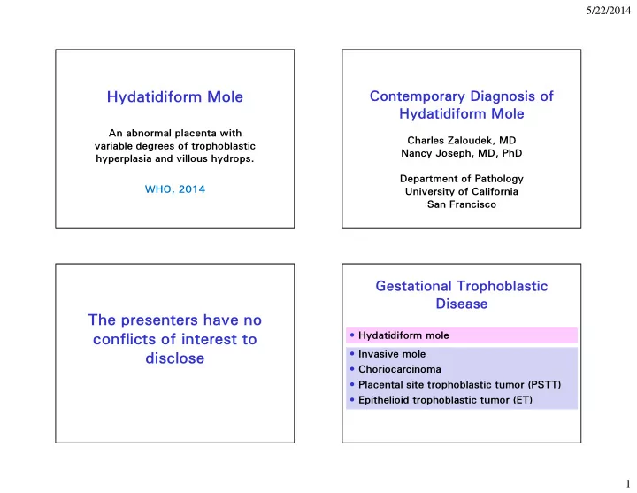
hydatidiform mole
play

Hydatidiform Mole Hydatidiform Mole An abnormal placenta with - PowerPoint PPT Presentation

5/22/2014 Contemporary Diagnosis of Hydatidiform Mole Hydatidiform Mole An abnormal placenta with Charles Zaloudek, MD variable degrees of trophoblastic Nancy Joseph, MD, PhD hyperplasia and villous hydrops. Department of Pathology WHO,


  1. 5/22/2014 Contemporary Diagnosis of Hydatidiform Mole Hydatidiform Mole An abnormal placenta with Charles Zaloudek, MD variable degrees of trophoblastic Nancy Joseph, MD, PhD hyperplasia and villous hydrops. Department of Pathology WHO, 2014 University of California San Francisco Gestational Trophoblastic Disease The presenters have no • Hydatidiform mole conflicts of interest to • Invasive mole disclose • Choriocarcinoma • Placental site trophoblastic tumor (PSTT) • Epithelioid trophoblastic tumor (ET) 1

  2. 5/22/2014 What is adequate study of a Specific Diagnosis of Gestations With Hydropic Villi Important POC Specimen? • TAB • Need to differentiate among – Probably reasonable to sign out on one – Complete hydatidiform mole slide with normal villi • Spontaneous/missed abortion – Partial hydatidiform mole – Must have a good view of villous – Hydropic abortion morphology • Different diseases – Suggest at least 2 slides with both fetal and maternal tissue • Different prognosis – One study found that it could require as • Different management many as 10 (!) slides to diagnose PM Complete Hydatiform Mole Scenario 1 Complete Mole 46XX “Homozygous” (80-90%) • Diploid • Androgenetic – all chromosomes are paternal * 46XpXp • Loss of maternally expressed imprinted transcripts, gain of paternally expressed imprinted transcripts 23X • 75-85% monospermic (46, XX) 46XX • 15-25% dispermic (46, XX or 46, XY) • Rare diploid biparental CHM * 2

  3. 5/22/2014 23X Complete Mole Changing Clinical Signs of Molar or Scenario 2 46XX 23Y “Heterozygous” (10-20%) Pregnancy or 46XY Incidence Then (16-17 wks) Now (< 12 wks) Vaginal bleeding 100 90 46XpXp Uterine enlargement 54 28 or T oxemia 22 1 46XpYp Hyperemesis 28 8 Hyperthyroidism 10 < 1 T rophoblastic emboli 2 < 1 46XX 23X Enlarged ovaries 15 15 or or No fetal heart sounds 100 100 46XY 23Y More likely to develop postmolar , 6 th Ed. Principles and Practice of Gynecologic Oncology trophoblastic neoplasms Complete Mole Ultrasound: Solid collection of echoes with numerous small anechoic spaces Ultrasound Diagnosis of Mole • Advanced cases of complete mole exhibit a characteristic vesicular pattern • In early cases cannot differentiate between complete mole and degenerating villi of abortion • Cystic villi in placenta and increased transverse diameter of gestational sac predict partial mole • In one recent study only 34% of moles suspected on ultrasound (58% complete, 17% partial) 3

  4. 5/22/2014 Traditional Complete Mole Histology • No fetal tissue • All villi abnormal • Rounded villous contours • Marked hydropic swelling • Central cisterns common • Vessels rare, no fetal RBC • Circumferential trophoblastic proliferation (CT, IT, ST) 4

  5. 5/22/2014 “Occasionally , the exuberance of the proliferating trophoblast taunts the pathologist into diagnosing choriocarcinoma. No matter how atypical the trophoblast is, nor how extensive the trophoblast hyperplasia, the presence of villous tissue, by definition, precludes a diagnosis of choriocarcinoma in a first or second-trimester placenta. Janice Lage, M.D. “Gestational T rophoblastic Diseases” in Robboy , Anderson, and Russell, Pathology of the Female Genital T ract Early Complete Hydatidiform Mole Early Complete Mole • Lack uniform hydropic change and diffuse circumferential trophoblastic hyperplasia. • Large bulbous or lobulated villi. • Basophilic villous stroma • Hypercellular villous stroma. • Karyorrhectic debris in villous stroma • Immature labyrinthine stromal vessels. • Focal circumferential trophoblastic hyperplasia. • Atypical IT in the implantation site. 5

  6. 5/22/2014 T rophoblastic Hyperplasia in Early Complete Hydatidiform Mole Hypercellular Villous Stroma in CHM Karyorrhectic Debris in Complete HM 6

  7. 5/22/2014 Atypical IT in complete mole implantation site Diploid Biparental Complete Mole • Very rare • Usually diagnosed when: – A patient has recurrent CHM – There is a familial tendency for CHM • Autosomal recessive inheritance • Mutations in a maternal gene involved in setting imprints in the ovum – 80% in NLRP7, a maternal gene on chromosome 19q – 5% in C6orf221 – 15% unknown Biparental CHM Partial Mole and Triploidy Can be similar to • 64 of 832 specimens triploid (~8%) androgenetic CHM (top) • 2/3 are diandric Can show less – Half, or 1/3 total were partial moles trophoblastic – Most of the remainder showed some but proliferation and not all features of a partial mole. budding (middle) • 1/3 are digynic Shows loss of p57 – None suspicious for partial mole staining Placenta 34:50-56, 2013 Human Pathol 29:505-51 1, 1998 7

  8. 5/22/2014 Partial Hydatiform Mole, Scenario 1: Dispermic – Heterozygous (Most Partial Mole Common – 90%) Clinical Findings • Most present with vaginal bleeding • Clinical diagnosis missed or incomplete 69, XmXpXp abortion 23X 69, XmXpYp • hCG titers rarely > 100,000mIU/ml 69, XmYpYp • Advanced complete mole type symptoms rare (preeclampsia, excessive uterine 23X or 23Y enlargement, theca-lutein cysts, hyperemesis, hyperthyroidism) Partial Hydatiform Mole, Scenario 2 Monospermic – Homozygous 10% 23X 69, XmXpYp 46 XY one sperm fertilizes the egg, followed by reduplication of the paternal chromosome set due to failure of meiosis I or II 8

  9. 5/22/2014 Partial Mole Histologic Features • Two populations of villi – Large hydropic villi with scalloped outlines – Normal to small fibrotic villi • Cisterns less conspicuous than in CM • Villi have inclusions • Trophoblastic proliferation moderate • Fetal parts or amnion may be present • Vessels and nucleated RBC may be present 9

  10. 5/22/2014 Invasive Mole • Persistent mole in which villi are: – Mainly in endometrium with early invasion of myometrium (“accreta pattern”) – Within myometrium, especially in blood vessels – Penetrate to serosa – Spread beyond uterus (vagina, lungs) Invasive Mole 10

  11. 5/22/2014 Invasive Mole Courtesy Michael Wells, MD Courtesy Kyu –Rae Kim, MD How well do pathologists diagnose hydatidiform mole? • Conran et al (AFIP, 1993) – Poor agreement with histology alone – Gross pathology data did not markedly improve concordance – Good agreement when flow cytometric data available • Crisp et al (Sheffield, 2003) – 40 cases; 2 revised on histology and 6 with special studies (20%) • Fukunaga et al (5 placental pathology experts, 2005) – Agreement of 4 or 5 in 30/50 cases – PHM vs HA main problem – With ploidy data agree in 39/50 cases – still problems with some cases of CHM vs HA • Vang et al (2012) – 80% accuracy for CHM with H&E, 96% with p57 – 78% accuracy for PHM 11

  12. 5/22/2014 Flow cytometry: Is it triploid or not? Ancillary Studies in the Diagnosis Complete mole or hydropic abortion of Hydatiform Mole • Cytogenetics Partial mole • FISH • Flow cytometry • Immunohistochemistry Complete mole, or, rarely, hydropic • Molecular genotyping abortion p57 kip2 • CDKN1C Gene - Cyclin dependent kinase inhibitor • Located on 11p15.5 • Paternally imprinted – expressed from maternal chromosome only • PHM and hydropic abortion – maternal present – Positive in cytotrophoblastic and stromal cells, in villous and extravillous intermediate trophoblastic cells and decidua – Negative in syncytiotrophoblastic cells • CHM – maternal absent – Negative in cytotrophoblastic, syncytiotrophoblastic, and stromal cells – Positive in villous and extravillous intermediate p57 partial HM trophoblastic cells and decidua 12

  13. 5/22/2014 p57 CHM p57 Almost Always Works • McConnell, T. G., et al. (2009). "Complete hydatidiform mole with retained maternal chromosomes 6 and 11." American Journal of Surgical Pathology 33 (9): 1409-1415. • Descipio, C., et al. (2011). "Diandric triploid hydatidiform mole with loss of maternal chromosome 11." American Journal of Surgical Pathology 35 (10): 1586-1591. Possible Hydatidiform Mole Gestational Trophoblastic Neoplasia p57 immunohistochemistry • In most patients treated for a mole, hCG returns to baseline within 60 days • GTN is persistent or recurrent trophoblastic Morphology c/w Flow Cytometry disease after treatment of a mole Complete Mole – 15-20 % after complete mole – 0.2-4 % after partial mole • Most likely diagnoses are persistent mole, Triploid Diploid invasive mole or choriocarcinoma • The clinician will treat the HCG titer and clinical features, not the diagnosis; a biopsy may not even be performed Complete Mole Partial Mole Non-molar 13

  14. 5/22/2014 Molecular Genotyping for Diagnosis of Hydatidiform Mole Is There a Way to Further Increase • Same cost as flow cytometry but faster turnaround Diagnostic Accuracy? • Provides ploidy data • Delineates parental source of Molecular Genotyping for Accurate chromosomes Diagnosis of Hydatidiform Moles – Dispermic vs monospermic – Diandric vs dygynic triploidy • Pioneered at Charing Cross, Yale, Johns Hopkins DNA contains microsatellites or Short Tandem Repeat (STR) short tandem repeats (STRs) 2-6 nucleotides • Short tandem repeats with repeating unit GT GT GT GT GT GT allele 1 of 2-6 base pairs • Distributed throughout the genome allele 2 • Non-coding DNA allele 3 • Heritable and stable allele 4 • Number of repeats is polymorphic (many different possible alleles at each locus) allele 5 allele 6 14

Download Presentation
Download Policy: The content available on the website is offered to you 'AS IS' for your personal information and use only. It cannot be commercialized, licensed, or distributed on other websites without prior consent from the author. To download a presentation, simply click this link. If you encounter any difficulties during the download process, it's possible that the publisher has removed the file from their server.

Recommend


More recommend