mdpi
play

MDPI http://sciforum.net/conference/mol2net-03 Antiproliferative - PDF document

MOL2NET , 2017 , 3, doi:10.3390/mol2net-03-04629 1 MOL2NET, International Conference Series on Multidisciplinary Sciences MDPI http://sciforum.net/conference/mol2net-03 Antiproliferative activity of Psidium guajava essential oil: a preliminary


  1. MOL2NET , 2017 , 3, doi:10.3390/mol2net-03-04629 1 MOL2NET, International Conference Series on Multidisciplinary Sciences MDPI http://sciforum.net/conference/mol2net-03 Antiproliferative activity of Psidium guajava essential oil: a preliminary study Matteo Radice a* , Matteo Chiurato b , Alessandra Guerrini b , Francesco Lozupone c a Universidad Estatal Amazónica, Km 2 ½ Via Napo (paso lateral), Puyo, Pastaza, Ecuador b Department of Life Sciences and Biotechnology (SVeB), UR7 Terra&Acqua Tech, University of Ferrara, Ferrara 44121, Italy; c Italian Center for Global Health. Italian National Institute of Health viale Regina Elena, 299 00161
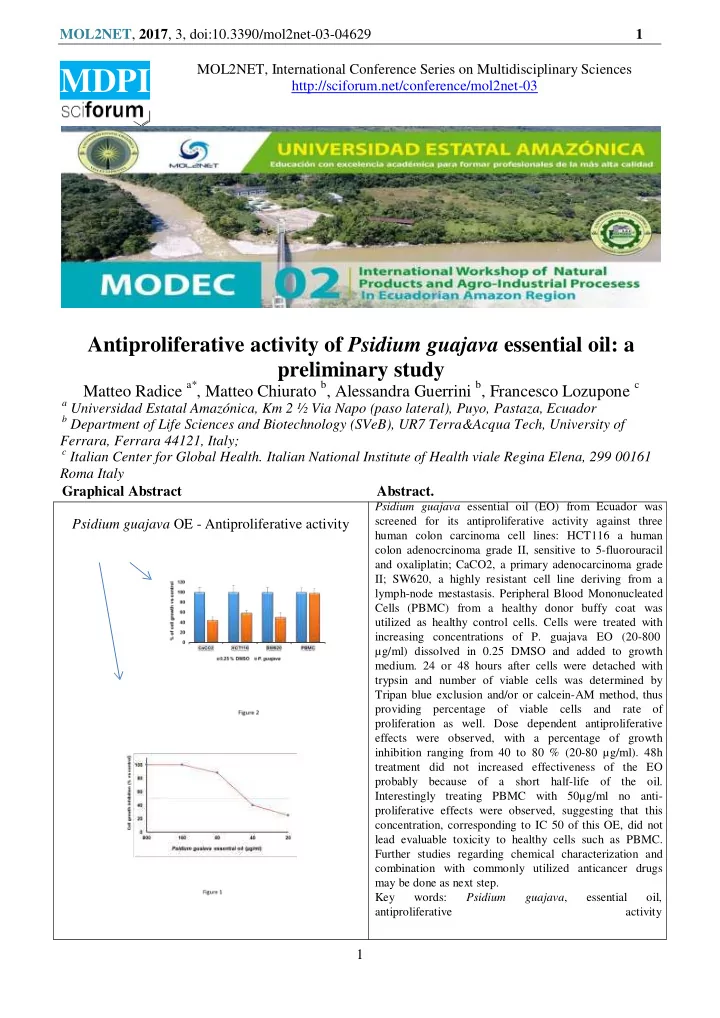
 Roma Italy Graphical Abstract Abstract. Psidium guajava essential oil (EO) from Ecuador was screened for its antiproliferative activity against three Psidium guajava OE - Antiproliferative activity human colon carcinoma cell lines: HCT116 a human colon adenocrcinoma grade II, sensitive to 5-fluorouracil and oxaliplatin; CaCO2, a primary adenocarcinoma grade II; SW620, a highly resistant cell line deriving from a lymph-node mestastasis. Peripheral Blood Mononucleated Cells (PBMC) from a healthy donor buffy coat was utilized as healthy control cells. Cells were treated with increasing concentrations of P. guajava EO (20-800 µg/ml) dissolved in 0.25 DMSO and added to growth medium. 24 or 48 hours after cells were detached with trypsin and number of viable cells was determined by Tripan blue exclusion and/or or calcein-AM method, thus providing percentage of viable cells and rate of proliferation as well. Dose dependent antiproliferative effects were observed, with a percentage of growth inhibition ranging from 40 to 80 % (20-80 µg/ml). 48h treatment did not increased effectiveness of the EO probably because of a short half-life of the oil. Interestingly treating PBMC with 50µg/ml no anti- proliferative effects were observed, suggesting that this concentration, corresponding to IC 50 of this OE, did not lead evaluable toxicity to healthy cells such as PBMC. Further studies regarding chemical characterization and combination with commonly utilized anticancer drugs may be done as next step. Key words: Psidium guajava , essential oil, antiproliferative activity 1

  2. MOL2NET , 2017 , 3, doi:10.3390/mol2net-03-04629 2 *Corresponding author: Matteo Radice - E-mail address: mradice@uea.edu.ec Introduction Psidium guajava is an important medicinal plant very well-known in several tropical countries, where extracts and metabolites of this plant are used in traditional medicine for the treatment of several diseases such as diabetes mellitus, diarrhea, dysentery, cardiovascular disorders and cancer. Many authors have reported traditional uses, folk medicine, phytochemistry and several studies demonstrating biological activities of P. guajava extracts [1-6]. Furthermore some papers suggest the anticancer potential of P. guajava leaf extracts by acting as inhibitors of tumor cells proliferation and motility and also acting as proapoptotic agents [7-10]. Differently from extracts, anti-proliferative effects of P. guajava essential oil (EO) is poorly characterized [11-12]. In this preliminary study, we investigated the antiproliferative activity of the P. guajava essential oil against human colon carcinoma cell lines. Materials and Methods Plant material Leaves of P. guajava were collected in the Amazonian region of Pastaza (Ecuador) and species authentication were certified by Dr. David Neill, voucher specimens were deposited at the Herbarium ECUAMZ of the Amazonian State University (UEA) in Ecuador (voucher specimen: Asanza 4814). The essential oil was obtained by hydrodistillation in a stainless steel distiller equipped wi th a Clevenger apparatus. Essential oil was obtained performing three distinct distillations and essential oil (moisture-free) yield was 0.14%. The oil was dried over anhydrous sodium sulphate and stored in sealed amber vials at 4°C. Cells PBMC: Peripheral Blood Mononucleated Cells (PBMC) were isolated from healthy donor’s buffy coats by gradient centrifugation. Colon cancer cell lines: CaCO2, a well differentiated grade II primary adenocarcinoma sensitive to 5-fluorouracil and oxaliplatin, HCT116 a malignant grade II colon adenocarcinoma and SW620, a highly resistant cell line derived from of a lymph-node mestastasis of grade III-IV colon adenocarcinoma patient. PMBC and cell lines were grown in RPMI1640 medium supplemented with 10% fetal bovine serum (FBS) and penicillin/streptomycin at 37 °C in a humidified atmosphere of 5% CO 2 . Antiproliferative activity : Cells were counted and plated in 24 multiwell plates at the same initial density. 24 after cell seeding, growth medium was replaced by fresh medium containing different concentrations of the oil dissolved in 0.25 % DMSO. 24 or 48 hours following P. guajava oil treatment (800 - 20 μ g/ml), cells were trypsinized, centrifugated, and resuspended in standard medium, and the anti-proliferative effect was quantified by performing a cell count with a hemocytometer. The cell viability was reported as a percentage of living cells compared to untreated control cells, by using the formula: Percentage of Living cells = Nb of treated cells/Nb of control cells × 100. Untreated control was considered as 100% living cells. Appropriate control groups with diluents only (0.25 % DMSO) and blank control were included. Cell viability was determined using the trypan blue (0.2% TB solution) exclusion test. The percentage of dead cells is calculated as (number of stained cells/number of total cells) × 100. P. guajava EO effects on viability or cytotoxicity were also evaluated by utilizing Calcein-AM/PI Double Stain Kit (Molecular Probes) according to the manufacturer protocol. 2

  3. MOL2NET , 2017 , 3, doi:10.3390/mol2net-03-04629 3 Results and Discussion As preliminary experiments, we setup a dose response curve to identify the range of oil concertation as optimal dose for the antitumor experiments. To address this issue we utilized HCT116 cells treated for 24 hours with different concentrations of P. guajava EO. Results shown in Figure 1 , allowed us to select the concentration of 50 µg/ml, roughly corresponding to the IC50 of the oil for these cells. Antiproliferative effects of P. guajava oil were then evaluated treating HCT116, SW480 and CaCO2 three different colon carcinoma cell lines for 24 or 48 hours with 50 μg /ml P. guajava oil in 0.25 % DMSO. As control 0.25 % DMSO was utilized, since no differences were observed between 0.25-1% DMSO medium and DMSO free RPMI. The results shown in Figure 2 clearly suggest the cell growth inhibitory effects of the oil in all cell lines tested that ranged from 50 to 60 %. 48h treatment did not increase the inhibitory effects in cell growth, probably because of a short half-life of the oil in the experimental conditions utilized (not shown). Furthermore no significant differences in cell viability between control and EO treated cells were observed, allowing us to hypothesize that cell growth inhibition, comes from antiproliferative, rather than cytotoxic effects. In these experiments, we assessed EO effects on Peripheral Blood Mononucleated Cells (PBMC) deriving from a healthy donor, to preliminarily assay potential toxic effects of oil; as shown in figure 2 P. guajava oil did not give any inhibitory or cytotoxic effects Conclusions In this manuscript, we describe for the first time anti-proliferative effects of P. guajava EO against colon cancer cells that represent one of the most aggressive human cancer. Although there is two papers about cytotoxic effects of this oil reviewed in [13], no data were available about its toxicity. Here we show the potential antitumor effects of P. guajava EO in an interval of concentration that is in the same range of several cytotoxic drugs already utilized for this type of tumors. Our observation about cytotoxicity, allowed us to hypothesize that this EO induces a cell growth arrest more than cytotoxicity, since did not verify significant differences in cell mortality. Interestingly we also observed that PBMC were unaffected by OE treatment, probably because of their state of resting, non- proliferating cells, suggesting that at the concentration utilized there are no toxic effects on healthy cells such as blood cells. From these encouraging results, we have planned to investigate the efficacy of these oils in combination with commonly utilized anticancer drugs. Next step will be the in vivo analysis of efficacy and toxicity, ad for the most promising oils the analysis of mechanism(s) of action in order to identify the target pathways. 3

Download Presentation
Download Policy: The content available on the website is offered to you 'AS IS' for your personal information and use only. It cannot be commercialized, licensed, or distributed on other websites without prior consent from the author. To download a presentation, simply click this link. If you encounter any difficulties during the download process, it's possible that the publisher has removed the file from their server.

Recommend


More recommend