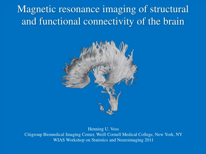

Magnetic resonance imaging of structural and functional connectivity of the brain Henning U. Voss Citigroup Biomedical Imaging Center, Weill Cornell Medical College, New York, NY WIAS Workshop on Statistics and Neuroimaging 2011
• Introduction • The MRI experiment • Diffusion tensor imaging, fiber orientation mapping, and neuronal fiber tracking • Functional connectivity: Resting state and optogenetic fMRI
Neuronal networks - from c. elegans to the human brain • C. elegans, exactly 302 neurons (959 cells total), 6393 synapses total • Small brains – Mouse ~16 × 10 6 neurons, ~ 8,000 synapses each. • Human brains – ~10 11 neurons, ~ 10,000 synapses each 3
Connectivity • C. elegans et al.: Optical imaging, electrical recordings, etc. • Small brains: In addition, tracers, brainbow, optogenetic fMRI. Example: mouseconnectome.org: 400 antero/retrograde tracer injections • Human brains: Limited in-vivo possibilities. Ex-vivo of limited use. Use DTI!
The MRI experiment
I(x,y,z) Image: Signal localization and Physics: “Spin echo” contrast mechanisms
Proton spins • For MRI, we are using the spin of atomic nuclei , mainly hydrogen nuclei • MRI does not affect Proton chemical processes and is noninvasive Electron • For protons and neutrons, spin = +/- 1/2 • For MRI, the atomic nuclei need to have a net spin and a charge to generate a magnetic moment • Good MR nuclei are 1 H , 13 C, 19 F, 23 Na, 31 P
Net magnetization • In a magnetic field B 0 the population ratio of spins parallel to B 0 versus spins anti-parallel to B 0 is roughly 100,006 to 100,000 (at room temperature) • Due to the surplus of aligned spins to non-aligned spins in an ensemble of spins, there is a small net magnetization (Bloch vector) M =(M x ,M y ,M z ) 8
Precession If M is not parallel to B , then it precesses clockwise around the direction of B. d M 1 1 ˆ Maxwell-Bloch equations: M B M M z M 0 z xy dt T T 1 2 Bloch vector Analogy: gyroscope 9
d M 1 1 ˆ M B M M z M 0 z xy dt T T 1 2 With B = (0,0,B 0 ) follows M xy (t)=M xy (0) exp(-i B 0 t) exp(-t/T 2 ) M z (t)=(1-exp(-t/T 1 )) M 0 /2p gyromagnetic ratio = 42.57 MHz/T B 0 main magnetic field [T]
Larmor equation: w 0 B 0 Constant gradients in object: w (x,y,z) = (B 0 +grad B * (x,y,z))
Generalize B = B 0 + G x (x,0,0): M xy (t) = M xy (0) exp( -i ( B 0 t+ G x x t)) exp(-t/T 2 ) =: M xy (0) exp( -i ( w 0 t+ k x x)) exp(-t/T 2 ) With magnetic field gradients the transverse magnetization looks like a spatial Fourier basis function
Fourier Transform Image space k-space y k y IFT x k x FT The MR signal is the 2D spatial Fourier transform of the imaged object. The image is the 2D inverse spatial Fourier transform of the k-space data
Diffusion weighted imaging (DWI)
“There is nothing that nuclear spins will not do for you, as long as you treat them as human beings" (Erwin L. Hahn 1949)
Theory o Rigorous approach: Add diffusion term to Bloch equations: Bloch-Torrey equation o More convenient approach: Start with probability distribution of spins and use diffusion equation
Bipolar pulsed gradient spin echo sequence (PGSE) 180º 90º Echo RF time TE Diffusion Gradients G G z R. Watts 2004
In one dimension: = length of diffusion gradients = spacing between diffusion gradients P(z) = probability distribution P(z 2 ,z 1 , ) = propagator: conditional probability that after a time the spins are at z 2 when they were at z 1 before
Assume << , then First gradient: dephasing 2nd gradient: dephasing Net dephasing (*)
Isotropic diffusion process: P ( z , z , t ) / t D P ( z , z , t ) (**) 2 1 2 1 P is Gaussian at t = : p 1 / 2 2 P ( z , z , ) ( 4 D ) exp( ( z z ) / 4 D ) 2 1 2 1 Combining this with (*), and using Einstein’s law <z 2 >=2Dt , we obtain (by integration in the complex domain) < 2 2 2 exp( ( ) ) exp( ( ) / 2 ) S G D G z Note that it is actually the diffusion path, not the diffusion constant, that is measured.
o We only consider lumped parameter model S = S 0 exp (-bD), where b is the b -value, fixed in experiment by Stejskal-Tanner equation: b = ( G 2 /3 o Example: D = 0.0007 mm 2 /s in in-vivo brain parenchyma b = 1/D = 1571 s/mm 2 as rule of thumb. Due to relaxation effects for finite diffusion gradient amplitudes (and therefore increased duration) smaller b = 800-1000 more appropriate for measuring ADC.
BUT: • Our assumptions of free, unlimited, isotropic Gaussian diffusion are not valid in the brain • One speaks of an apparent diffusion constant or ADC This is good!
Diffusion tensor imaging
Anisotropic attenuation o Remember: One dimensional lumped parameter model: S = S 0 exp (-bD) o Now: Directionality dependence S = S 0 exp (- b g t D g ), where g is a vector containing the diffusion gradient direction, and D is the diffusion tensor
180º 90º Echo RF time TE G x G y G z R. Watts 2004
Westin et al. 2002
The diffusion tensor Jellison 2004
Some invariants (not depending on angle of coordinate system): Mean diffusivity (ADC) = tr(D)/3 = (D xx +D yy +D zz )/3 Fractional anisotropy (FA) =
What is measured? Restricted, permeable barrier, hindered diffusion? Le Bihan 1995
Interpreting ADC and FA Diffusivity and FA are related to the density , size, type, and myelination of fibers. Myelination of fibers Number of fibers High Diffusivity High FA High Diffusivity High FA Low Diffusivity Low FA Low Diffusivity Low FA Size of fibers Directionality of Fibers High FA Low FA High FA High Diffusivity Low Diffusivity Same Diffusivity Same Diffusivity Low FA S. Maier and M. Kubicki http://www.na-mic.org/
How many measurements? o One needs to measure the symmetric diffusion tensor and a b=0 weighted image o 6+1=7 measurements o It has been shown that more = better: o 6 icosahedral directions are not rotationally invariant (precision matrix contains 15 independent parameters and depends on tensor itself)
Fiber orientation mapping
Intensity = Fractional anisotropy Color = main fiber direction:
White matter fiber tractography
Direction of greatest diffusion R. Watts 2004, Mori et al, 1999
Application: Presurgical planning Neurosurgery/Radiology at NYPH (tumor & epileptic surgery) and CBIC
39
Non-Gaussian diffusion
q-space imaging Resolves intravoxel orientational heterogeneity (partial voluming of different fiber tracts and crossing / branching / kissing fibers)
o Remember: o Define q = gd G and z = z 2 -z 1 to get o Therefore, the signal S is again the Fourier-transform of a density P o By inversion, one can measure P. o This requires lots of q -values, i.e., one needs to vary timing d or gradient strength G .
Q-ball imaging o Drastically reduced scan time o HARDI-sequence (High angular resolution diffusion imaging) o Spherically sampled data o Postprocessing (Funk Radon transform)
The orientation distribution function (ODF) ( r and q are reciprocal vectors) ODF:
Q-Ball imaging Sample only on a sphere, not on 3D volume: Funk-Radon transform = extension of planar Radon transform to the sphere = transform from sphere to sphere = line integral along equator of sphere, for each vector on sphere w = unit direction vector f ( w ) = scalar function on sphere u = direction of interest
Extended FRT: Maps from 3D Cartesian space to sphere x = unit direction vector r = particular radius at which FRT is evaluated f ( x ) = scalar function in 3D Cartesian space
Theorem (D. S. Tuch): The extended FRT of the diffusion signal gives a strong approximation to the ODF, i.e., ( u and q are reciprocal vectors) The sum of the diffusion signal over an equator approximately gives the diffusion probability in the direction normal to the plane of the equator.
Central element: The diffusion orientation distribution function (ODF) (a) Diffusion signal sampled on fivefold tessellated icosahedron ( m 252). The signal intensity is indicated by the size and color (white yellow red) of the dots on the sphere. ( b ) Regridding of diffusion signal onto set of equators around vertices of fivefold tessellated dodecahedron. ( c ) Diffusion ODF calculated using FRT. ( d ) Color-coded spherical polar plot rendering of ODF. ( e ) Min – max normalized ODF. (Tuch 2004)
ODF map of caudal midbrain cp, cerebral peduncle; ctt, central tegmental tract; fp, frontopontine tract; rst, reticulospinal tract; scp, superior cerebellar peduncle; sn, substantia nigra; xscp, crossing of the superior cerebellar peduncle.
Recommend
More recommend