Functional Magnetic Resonance Imaging Gregory Hong Baiheng Xu 2 - - PowerPoint PPT Presentation

functional magnetic resonance imaging
SMART_READER_LITE
LIVE PREVIEW

Functional Magnetic Resonance Imaging Gregory Hong Baiheng Xu 2 - - PowerPoint PPT Presentation

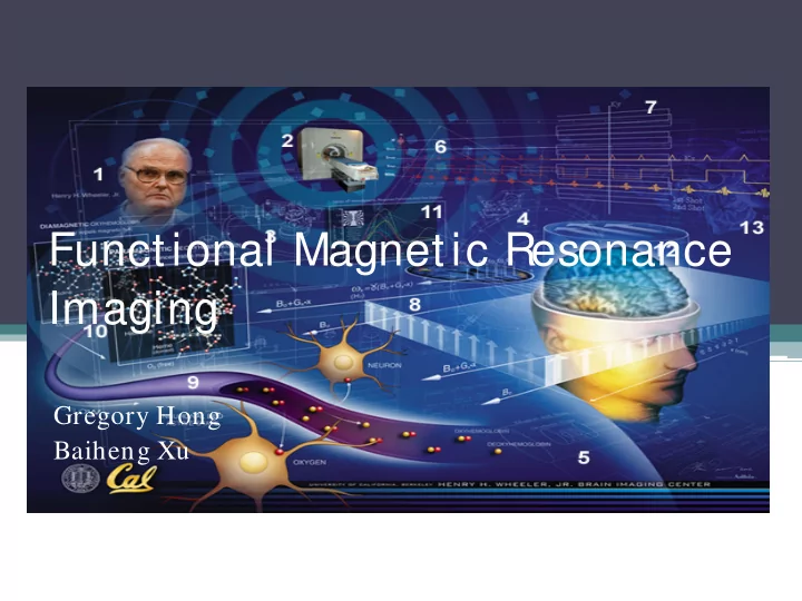
Functional Magnetic Resonance Imaging Gregory Hong Baiheng Xu 2 What is fMRI Functional magnetic resonance imaging, or fMRI, is a technique for measuring brain activity. It works by detecting the changes in blood oxygenation and flow


slide-1
SLIDE 1

Functional Magnetic Resonance Imaging

Gregory Hong Baiheng Xu

slide-2
SLIDE 2

What is fMRI

  • Functional magnetic resonance imaging, or fMRI, is a technique for

measuring brain activity.

  • It works by detecting the changes in blood oxygenation and flow

that occur in response to neural activity – when a brain area is more active it consumes more oxygen and to meet this increased demand blood flow increases to the active area.

2

slide-3
SLIDE 3

MRI

3

slide-4
SLIDE 4

fMRI

4

slide-5
SLIDE 5

S canner

5

slide-6
SLIDE 6

How MRI works

6

slide-7
SLIDE 7

What does MRI Measure?

7

slide-8
SLIDE 8

What does fMRI Measure?

  • Oxygen is delivered to neurons by haemoglobin in capillary

red blood cells.

  • Haemoglobin is diamagnetic when oxygenated
  • Haemoglobin is paramagnetic when deoxygenated.
  • Blood oxygenation level dependent (BOLD) imaging.

8

slide-9
SLIDE 9

What does fMRI Measure?

9

slide-10
SLIDE 10

fMRI… What is it good for?

  • fMRI is used to monitor brain functions of both

healthy and ailing individuals

  • Used on healthy individuals to study and

understand brain anatomy and cognition

  • Used on ill individuals to diagnose neurological

problems as well as pre-surgical diagnosis and risk analysis

10

slide-11
SLIDE 11

fMRI as a diagnostic tool

  • BOLD fMRI is used a part of pre-surgical

procedure to identify risks and to plan for the performance of aggressive procedures to the central region of the brain

  • Also has a role in identifying patients who are

not ideal candidates for surgery and should opt for less invasive therapeutic methods

11

slide-12
SLIDE 12

BOLD fMRI for Brain Tumor S urgery

12

slide-13
SLIDE 13

What the picture really looks like

13

slide-14
SLIDE 14

fMRI in cognitive research

  • fMRI is used to map the brain in order to

understand brain function during cognitive activity

  • An example of this is a study done at the

Stanford School of Medicine into brain behavior while listening to classical music

14

slide-15
SLIDE 15

This is your brain on music

http://med.stanford.edu/news_releases/2007/july/music.html 15

slide-16
SLIDE 16

History: NMR spectroscopy

  • Nuclear magnetic resonance (NMR): precursor

to MRI technology

  • Effect: They found that when certain naturally-
  • ccurring nuclei were placed in a magnetic field

they absorbed energy in the radio frequency range of the electromagnetic spectrum.

  • Used extensively in chemistry
  • Felix Bloch and Edward Purcell

won the 1952 Nobel Prize in Physics

16

slide-17
SLIDE 17

History: The beginnings of MRI

  • Feb. 9, 1974: Dr. Raymond V. Damadian patents

a method for distinguishing normal from cancerous tissue by what was then called nuclear magnetic resonance.

  • 1977: First MRI exam performed on human

patient; procedure long and complicated

  • March 17, 1985: New York Times runs an article

explaining that most doctors were calling both the procedure and the machines “magnetic resonance imaging”

17

slide-18
SLIDE 18

History: Advancing MRI

  • 1973: Paul Lauterbur described his research on the high

level of contrast that could be realized in nuclear magnetic resonance imaging

  • Sir Peter Mansfield developed methodology to analyze

the signals and assemble them rapidly into three- dimensional images.

18

slide-19
SLIDE 19

History: The dawn of fMRI

  • BOLD fMRI arose from the discovery that there is a

change in blood oxygenation levels with brain activity by Seiji Ogawa, who reported his team’s findings in 1990

  • Robert Turner then showed the utility of blood
  • xygenation characteristics by using cats with induced

hypoxia (inadequate oxygen supply)

  • The first movies of human visual cortex activation were

produced at the Massachusets General Hospital in May

  • f 1991
  • Perfusion fMRI uses measurements of blood flow in the

brain to determine brain activity; Dr. John Detre published his paper on this subject in 1992.

19

slide-20
SLIDE 20

fMRI T1/ T2 Mechanism

  • The two fundamental MRI relaxation rates T1 and

T2 are used to describe fMRI signal

  • T1 is the rate at which nuclei approach thermal

equilibrium, and is used to measure perfusion

  • T2 is more important to BOLD as it represents the

rate of decay of MRI signal due to magnetic field inhomogeneities, and thus can be used to measure blood oxygenation change

  • An observed increased signal on T2 weighted images

during activation reflects a decrease in deoxyhemoglobin content

20

slide-21
SLIDE 21

S ignal Intensity of BOLD fMRI

21

slide-22
SLIDE 22

22

slide-23
SLIDE 23

The current state of fMRI

  • Current MRI systems can use BOLD techniques

to acquire whole head images with a spatial resolution of 1.5x1.5 mm in plane and 3 mm slice thickness

  • “Siemens MAGNETOM Verio is the first MRI

system to offer 3T field strength combined with a 70cm bore and Tim (Total imaging matrix) technology” (Siemens fMRI brochure)

23

slide-24
SLIDE 24

fMRI software

  • Siemens MRI machines can utilize their proprietary

syngo Neuro Suite

  • “It includes techniques such as single-shot epi (echo

planar imaging) as well as MPRAGE used to acquire the anatomic information that will later be fused with the fMRI data.”

24

slide-25
SLIDE 25

Advantages

  • No radiation.
  • No injection.
  • High spatial resolution. Typical 2–3 mm,

1mm possible.

  • Oversee all regions of the brain.
  • Compelling images of brain “activation”

regions.

25

slide-26
SLIDE 26

Disadvantages

  • Expensive
  • Influence by non-neural changes in the body.
  • Poor temporal resolution. The BOLD response peaks

approximately 5 seconds after neuronal firing begins in an area. EEG have higher temporal resolution but worse spatial resolution.

  • Minimizing the distributed nature of processing in neural

networks.

  • The BOLD response can be affected by a variety of

factors, including: drugs, age, brain pathology, attention, amount of carbon dioxide in the blood.

26

slide-27
SLIDE 27

Limitations

  • High-quality images are assured only person remain

perfectly still

  • A person who is very large may not fit.
  • MRI generally is not recommended for patients who

have been acutely injured.

  • Examination takes Long time.
  • Pregnant women are advised not to have an MRI

exam unless medically necessary.

  • Cost
  • Additional tests for confirmation is required.

27

slide-28
SLIDE 28

Future

slide-29
SLIDE 29

Read your mind

29

slide-30
SLIDE 30

The Future of Mind Reading

30

slide-31
SLIDE 31

Questions?

31

slide-32
SLIDE 32

References

  • Bakalar, Nikolas. "M.R.I. 1974." The New York Times 17 May 2011, New York ed., D7
  • sec. New York Times. 16 May 2011. 27 Nov. 2011

<http:/ / www.nytimes.com/ 2011/ 05/ 17/ health/ 17first.html?_r=4>.

  • Baker, Mitzi. "Music moves brain to pay attention, Stanford study finds." Stanford

School of Medicine News Releases. 1 Aug. 2007. Stanford School of Medicine. 27

  • Nov. 2011 <http:/ / med.stanford.edu/ news_releases/ 2007/ july/ music.html>.
  • "Brain Imaging, functional (fMRI)." RadiologyInfo - The radiology information

resource for patients. 27 Nov. 2011 <http:/ / www.radiologyinfo.org/ en/ info.cfm?pg=fmribrain>.

  • "The Future Role of functional MRI in Medical Applications." About functional MRI.

Program for Imaging and Cognitive Sciences, Columbia University. 27 Nov. 2011 <http:/ / www.fmri.org/ fmri.htm>.

  • "Historical Perspective of Biomedical Imaging: From MRI to fMRI." NIBIB: Stories
  • f Discovery. 25 June 2009. National Institude of Biomedical Imaging and
  • Bioengineering. 27 Nov. 2011

<http:/ / www.nibib.nih.gov/ HealthEdu/ Discovery/ HistPerspective>.

  • Kwong, Kenneth K., and David A. Chesler. "Chapter 12.3: Functional MRI." Medical

devices and systems. By Joseph D. Bronzino. Boca Raton, FL: CRC/ Taylor & Francis, 2006.

32

slide-33
SLIDE 33

References Con’ t.

  • "The Nobel Prize in Physiology or Medicine 2003." Nobelprize.org. 27 Nov. 2011

<http:/ / www.nobelprize.org/ nobel_prizes/ medicine/ laureates/ 2003/ >.

  • Siemens. Delivering the most exciting equation in MRI. MAGNETOM Verio.
  • Brochure. Author, 2008. 27 Nov. 2011

<http:/ / www.medical.siemens.com/ siemens/ en_US/ gg_mr_FBAs/ files/ brochures/ Verio_brochures/ brochure_Verio_en_07-2008.pdf>.

  • Siemens. Functional Magnetic Resonance Imaging (fMRI). Brochure. Author, 2008.

27 Nov. 2011 <http:/ / www.medical.siemens.com/ siemens/ en_GB/ gg_mr_FBAs/ files/ MAGNETO M_World/ MW_MRI_HotTopics/ fMRI.pdf>.

  • Stippich, Christoph. "Chapter 7: Preoperative Blood Oxygen Level Dependent

(BOLD) functional Magnetic Resonance Imaging (fMRI) of Motor and Somatosensory Function." FMRI Basics and Clinical Applications. By Stephan Ulmer and Olav Jansen. Berlin: Springer, 2010. 51-68.

33

slide-34
SLIDE 34

References: Images and Videos

  • http:/ / www.youtube.com/ watch?v=8jc8URRxPIg
  • http:/ / www.youtube.com/ watch?v=9E1GoWhSlho&

feature=related

  • http:/ / www.youtube.com/ watch?v=e0tQY4_UUew
  • http:/ / blogs.oem.indiana.edu/ scholarships/ index.p

hp/ 2009/ 10/ 26/ neurons-and- electrodes/ fmri_groot/

  • http:/ / neurolove.tumblr.com/ post/ 789433845/ mri
  • magnetic-resonance-imaging-so-what-is
  • http:/ / www.wcaslab.com/ tech/ plga.htm
  • http:/ / www.youtube.com/ watch?v=VaQ66lDZ-08

34